


|
Trauma X-ray - Axial skeleton Cervical spine - Normal anatomy. C-spine systematic approach - Normal Lateral 1. C-spine systematic approach - Normal Lateral 2. C-spine normal anatomy - Lateral (detail). C-spine systematic approach - Normal AP. Odontoid peg/Open mouth view. C-spine normal anatomy - Open mouth view. Open mouth view -. Rotated. . C-spine normal anatomy - ' Swimmer's' view. C-spine - Systematic approach. C-spine systematic approach - Normal Lateral 1. C-spine systematic approach - Normal Lateral 2. C-spine normal anatomy - Lateral (detail). C-spine systematic approach - Normal AP. C-spine normal anatomy - Open mouth view. Open mouth view - Rotated. C-spine normal anatomy - ' Swimmer's' view. Show
Top 1: Trauma X-ray - Axial skeleton - Cervical spine - Normal anatomyAuthor: radiologymasterclass.co.uk - 168 Rating
Description: Trauma X-ray - Axial skeleton Cervical spine - Normal anatomy. C-spine systematic approach - Normal Lateral 1. C-spine systematic approach - Normal Lateral 2. C-spine normal anatomy - Lateral (detail). C-spine systematic approach - Normal AP. Odontoid peg/Open mouth view. C-spine normal anatomy - Open mouth view. Open mouth view -. Rotated C-spine normal anatomy - ' Swimmer's' view. C-spine - Systematic approach. C-spine systematic approach - Normal Lateral 1. C-spine systematic approach - Normal Lateral 2. C-spine normal anatomy - Lateral (detail). C-spine systematic approach - Normal AP. C-spine normal anatomy - Open mouth view. Open mouth view - Rotated. C-spine normal anatomy - ' Swimmer's' view.
Matching search results: Cervical spine anatomy - X-ray appearances. Normal c-spine x-ray. Lateral c-spine x-ray description. Systematic approach to cervical spine x-ray interpretation. AP cervical spine x-ray appearances. Odontoid peg view description. Odontoid peg view - open mouth view - X-ray. Swimmer view X-ray of the cervico-thoracic junction. ...
Top 2: Chest (lateral decubitus view) | Radiology Reference Article ...Author: radiopaedia.org - 125 Rating
Description: Citation, DOI & article data. Related articles: Imaging in practice . Promoted articles (advertising) Citation, DOI & article dataCitation:Murphy, A. Chest (lateral decubitus view). Reference article, Radiopaedia.org. (accessed on 13 Nov 2022) https://doi.org/10.53347/rID-53650The lateral decubitus view of the chest is a specialized projection that is now rarely used due to the ubiquity of CT. It is chiefly used in the pediatric. population.On this page:Undertaken to demonstrate small
Matching search results: Sep 19, 2021 · 35 cm x 43 cm; exposure. 100 - 125 kVp; 3 - 10 mAs; SID. 100 cm; grid. yes; Image technical evaluation. A marker annotating 'horizontal beam decubitus" should always be present, with the side of interest clearly labeled. The entire lungs should be visible from the apices down to the lateral costophrenic angles. the chin should not be ... ...
Top 3: A paediatric X-ray exposure chart - PMC - National Center for ...Author: ncbi.nlm.nih.gov - 118 Rating
Description: Exposure groups based on patient size variability. Kilovoltage peak manipulation. Additional beam filtration. Detector exposure, automatic exposure control, and mAs manipulation. Antiscatter grid selection. Image processing technologies Journal List J Med Radiat Sci v.61(3); 2014 Sep PMC4175850 J Med Radiat Sci. 2014 Sep; 61(3): 191–201. AbstractThe aim of this review was to develop a radiographic optimisation strategy to make use of digital radiography (DR) and needle phosphor computerised ra
Matching search results: Jun 09, 2014 · It is recommended that purchasing digital X-ray equipment with high detective quantum efficiency detectors, and then optimising the exposure chart for use with these detectors is of high importance for sites performing paediatric imaging. ... Lateral cervical spines are taken at 150 cm. Erect chest X-rays are taken at 180 cm. Full leg/full ... ...
 Top 4: Cervical Spine Injury - RCEMLearningAuthor: rcemlearning.co.uk - 99 Rating
Description: (a) Plain cervical spine series. (b). Swimmers view (see Figures 9 and 10):. (c) Flexion / extension. views. (d) Computerised Tomography (CT). (e) Magnetic Resonance Imaging (MRI) Patients, whose cervical spine cannot be clinically cleared, will require imaging to permit identification or exclusion of significant injury.(a) Plain cervical spine seriesPlain radiographs do not detect all cervical spine fractures. In 29 published studies 16% of fractures (range 0 to 67%) were missed11. Plain lateral
Matching search results: Oct 11, 2021 · Figure 8: Normal series of plain cervical spine x-rays (i) Lateral view: ... Figure 10(a): Inadequate plain lateral x-ray. Figure 10(b): Swimmers view now demonstrating C7/T1 (c) Flexion / extension views. For many years flexion/extension views used to be recommended for the clearing of the cervical spine. A number of studies have demonstrated ... ...
 Top 5: Browse Articles | NatureAuthor: nature.com - 62 Rating
Description: Saying ‘no’ in science isn’t enough. The neurons that restore walking after paralysis Article Type All Article (21463). Book Review (21064) Books & Arts (2987). Books Received (28076) Correspondence (11692). Editorial (4066) Erratum (1641). Letter (120287) Matters Arising (1672). Miscellany (7852) News (103867). News & Views (40213) News Feature (2267). News in Brief (1760) Obituary (6978). Opinion (5080) Research Highlight (2051). Research Highlights (4493) Scientific Correspondence (4028). Su
Matching search results: Nov 09, 2022 · Fresh data released at the climate summit show global carbon dioxide emissions from fossil fuels are soaring despite energy crisis. ...
 Top 6: Thorax - WikipediaAuthor: en.wikipedia.org - 54 Rating
Description: Human thorax images[edit]. Clinical significance[edit]. Anatomical. landmarks[edit]. Non-cardiac causes of chest pain[edit] . ThoraxChestX-ray image of the chest showing the. internal anatomy of the rib cage, lungs and heart as well as the inferior thoracic border–made up of the diaphragm.. Surface projections of the organs of the trunk, with the thorax or chest region seen stretching down to approximately the end of the. oblique lung fissure anteriorly, but more deeply its lower limit rather corres
Matching search results: The thorax or chest is a part of the anatomy of humans, mammals, and other tetrapod animals located between the neck and the abdomen. In insects, crustaceans, and the extinct trilobites, the thorax is one of the three main divisions of the creature's body, each of which is in turn composed of multiple segments.. The human thorax includes the thoracic cavity and the thoracic wall. ...
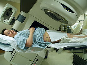 Top 7: Radiation therapy - WikipediaAuthor: en.wikipedia.org - 76 Rating
Description: Use in non-cancerous diseases[edit]. Further. reading[edit]. Acute side effects[edit]. Late side effects[edit]. Cumulative side effects[edit]. Effects on reproduction[edit]. Effects on pituitary system[edit]. Radiation therapy. accidents[edit]. Mechanism of action[edit]. External beam radiation. therapy[edit]. Contact X-ray. brachytherapy[edit]. Brachytherapy (sealed source. radiotherapy)[edit]. Radionuclide therapy[edit]. Intraoperative. radiotherapy[edit]. Schedules for. fractionation[edit]. Estimation of dose based on target sensitivity[edit]. Conventional external beam radiation. therapy[edit]. Stereotactic. radiation[edit]. Virtual simulation, and 3-dimensional conformal radiation therapy[edit]. Intensity-modulated radiation therapy. (IMRT)[edit]. Volumetric modulated arc therapy (VMAT)[edit]. Temporally feathered radiation therapy (TFRT)[edit]. Automated planning[edit]. Particle therapy[edit]. Motion. compensation[edit].
Matching search results: Radiation therapy or radiotherapy, often abbreviated RT, RTx, or XRT, is a therapy using ionizing radiation, generally provided as part of cancer treatment to control or kill malignant cells and normally delivered by a linear accelerator.Radiation therapy may be curative in a number of types of cancer if they are localized to one area of the body. It may also be used as part of … ...
 Top 8: Home Page: Journal of Manipulative & Physiological TherapeuticsAuthor: jmptonline.org - 90 Rating
Description: Most Read (Last 30 Days). Most Cited (Previous 3 Years). Journal. of Manipulative & Physiological Therapeutics. National University of Health Sciences MastheadCurrent IssueJournal of Manipulative & Physiological TherapeuticsArticles in PressIssue HighlightsAboutJournal. of Manipulative & Physiological TherapeuticsThe Journal of Manipulative and Physiological Therapeutics (JMPT) is. an international and interdisciplinary journal dedicated to the advancement of conservative health care princ
Matching search results: Nov 03, 2022 · Journal of Manipulative & Physiological Therapeutics now recommends you submit all manuscripts electronically. Submit Manuscript. You may also use this system to track your manuscript through the review process. ...
 Top 9: Normal lateral cervical spine radiograph | Radiology CaseAuthor: radiopaedia.org - 135 Rating
Description: Citation, DOI & case data. 12 public playlists include this. case. Related Radiopaedia. articles. Promoted articles (advertising) Case contributed by Dr Ian Bickle ◉Diagnosis certain Diagnosis certain . Add to Report problem with CaseContact userCitation, DOI & case data. Citation:Bickle, I. Normal lateral cervical spine radiograph. Case study, Radiopaedia.org. (accessed on 13 Nov 2022) https://doi.org/10.53347/rID-46395DOI:https://doi.org/10.53347/rID-46395Permalink:https://rad
Matching search results: Jul 4, 2016 · Normal plain radiograph of lateral cervical spine. 12 public playlists include this case.Jul 4, 2016 · Normal plain radiograph of lateral cervical spine. 12 public playlists include this case. ...
 Top 10: Normal cervical spine radiographs | Radiology Case - RadiopaediaAuthor: radiopaedia.org - 135 Rating
Description: Citation, DOI & case data. Scroll to see annotations. Scroll to see lines and measurements. 3 articles feature images from this case. 67 public playlists include this case. Related Radiopaedia articles. Promoted articles (advertising) Case contributed by Dr Andrew Dixon ◉Diagnosis not applicable Diagnosis not applicable . Add to Report problem with CaseContact userCitation, DOI & case data. Citation:Dixon, A. Normal cervical spine radiographs. Case study, Radiopaedia.org. (acc
Matching search results: Nov 29, 2014 · Normal cervical spine radiographs in a young adult. ... Normal lateral cervical spine lines and measurements.Nov 29, 2014 · Normal cervical spine radiographs in a young adult. ... Normal lateral cervical spine lines and measurements. ...
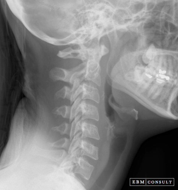 Top 11: Lateral Cervical Spine Radiograph (X-Ray) - How to ReadAuthor: ebmconsult.com - 141 Rating
Description: Basic Anatomy & Lines Note:Scroll over or tap on image to see labels & lines. Note: Scroll over or tap on image to see labels & lines. Core Radiographic Lines. Note: Scroll over or tap on the image to see labels & lines Note: Scroll over or tap on the image to see labels & lines . McRae Line:This is a line drawn on a lateral radiograph of the skull or on a sagittal cut from a CT or MRI scan that connects the posterior and anterior aspects of the foramen magnum. The t
Matching search results: Note: If the tip of the dens is eroded then the Redlund-Johnell and modified Ranawat methods (normal CT values for men is > 23.7 mm and for women is > 24.2 mm) ...Note: If the tip of the dens is eroded then the Redlund-Johnell and modified Ranawat methods (normal CT values for men is > 23.7 mm and for women is > 24.2 mm) ... ...
Top 12: Trauma X-ray - Axial skeleton - Cervical spine - Normal anatomyAuthor: radiologymasterclass.co.uk - 168 Rating
Description: Trauma X-ray - Axial skeleton Cervical spine - Normal anatomy. C-spine systematic approach - Normal Lateral 1. C-spine systematic approach - Normal Lateral 2. C-spine normal anatomy - Lateral (detail). C-spine systematic approach - Normal AP. Odontoid peg/Open mouth view. C-spine normal anatomy - Open mouth view. Open mouth view -. Rotated C-spine normal anatomy - ' Swimmer's' view. C-spine - Systematic approach. C-spine systematic approach - Normal Lateral 1. C-spine systematic approach - Normal Lateral 2. C-spine normal anatomy - Lateral (detail). C-spine systematic approach - Normal AP. C-spine normal anatomy - Open mouth view. Open mouth view - Rotated. C-spine normal anatomy - ' Swimmer's' view.
Matching search results: The 3 standard views are - Lateral view - Anterior-Posterior (AP) view - and the Odontoid Peg view (or Open Mouth view). In the context of trauma these images ...The 3 standard views are - Lateral view - Anterior-Posterior (AP) view - and the Odontoid Peg view (or Open Mouth view). In the context of trauma these images ... ...
 Top 13: Head and Neck – Undergraduate Diagnostic Imaging FundamentalsAuthor: pressbooks.pub - 125 Rating
Description: The following are normal x-rays of the cervical spine (C-spine):Posterior-AnteriorLateral. Oblique viewNormal Adult C-Spine, LabelledAP Adult C-spine, LabelledLateral Adult C-spine, LabelledSwimmer’s. View Adult C-spine, LabelledOblique View Adult C-spine, LabelledOpen Mouth, Odontoid, Adult C-spine, LabelledThe following are normal images of the Pediatric C-Spine, unlabelled and labelled:The following are normal CT images of the brain:The ODIN link provides the full set of. images and they can be
Matching search results: The following are normal x-rays of the cervical spine (C-spine):. Posterior-Anterior. Lateral ... AP Adult C-spine, Labelled Lateral Adult C-spine, Labelled.The following are normal x-rays of the cervical spine (C-spine):. Posterior-Anterior. Lateral ... AP Adult C-spine, Labelled Lateral Adult C-spine, Labelled. ...
 Top 14: Radiographic Comparison between Cervical Spine Lateral ... - NCBIAuthor: ncbi.nlm.nih.gov - 118 Rating
Description: Materials and Methods. Radiographic Measurement Journal List Global Spine J v.6(2); 2016 Mar PMC4771508 Global Spine J. 2016 Mar; 6(2): 118–123. Abstract Study Design Retrospective radiologic study. Objective The sagittal alignment of the cervical spine can be evaluated using either a lateral cervical radiograph or a whole-spine lateral view on which the cervical spine is included. To our. knowledge, however, no report has compared the two. The purpose of this work is to identify the difference
Matching search results: Jun 24, 2015 · Normal lordotic angles for C2–C7 have been reported to range from 20 to 35 degrees, but these values are highly related to the method of ...Jun 24, 2015 · Normal lordotic angles for C2–C7 have been reported to range from 20 to 35 degrees, but these values are highly related to the method of ... ...
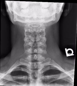
 Top 15: Cervical Spine X-ray Interpretation - OSCE Guide - Geeky MedicsAuthor: geekymedics.com - 134 Rating
Description: Acquire all necessary views. Cartilage (i.e discs). Antero-posterior (AP) view. Odontoid/open-mouth view Cervical spine X-rays aren’t something you’ll see commonly outside of an Emergency Department, but the importance of being able to read them, and the risks of missing significant findings (spinal cord injury, death) make brushing up on the basics well worthwhile! It is worth noting though, that if you are in any doubt regarding your interpretation of these often-difficult images, ask for hel
Matching search results: Nov 12, 2021 · Pre-vertebral (i.e the area directly anterior to the vertebral bodies) soft tissue is best assessed using a lateral view. Soft tissue appears as ...Nov 12, 2021 · Pre-vertebral (i.e the area directly anterior to the vertebral bodies) soft tissue is best assessed using a lateral view. Soft tissue appears as ... ...
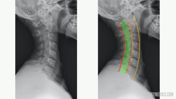
 Top 16: C-spine x-ray interpretation - Don't Forget the BubblesAuthor: dontforgetthebubbles.com - 117 Rating
Description: The ABC’s of the cervical spine provide a helpful mnemonic to guide the systematic assessment of these x-rays. Remember; you require all three views (lateral, AP and odontoid/open mouth view) for an adequate study.A: Adequacy. The C7/T1 junction must be visibleA: Alignment. Ensure. all 4 lines are contiguous/uninterrupted 1. Anterior longitudinal line 2. Posterior longitudinal line 3. Spinolaminal line 4. Spinous process lineB: Bones. Each vertebrae must be examined for fracture/co
Matching search results: Dec 11, 2017 · C-spine x-ray interpretation, Don't Forget the Bubbles, 2017. ... Normal <3 mm; >3 mm (XR) or 2 mm (CT) ?damage to transverse ligament ...Dec 11, 2017 · C-spine x-ray interpretation, Don't Forget the Bubbles, 2017. ... Normal <3 mm; >3 mm (XR) or 2 mm (CT) ?damage to transverse ligament ... ...
|

Top 16 normal lateral cervical x ray 2022


Copyright © 2024 boxhoidap Inc.