


Background Show
Gallbladder anatomy (overview).
Clinical FeaturesDifferential DiagnosisRUQ Pain
Workup
Management
Disposition
See Also
External LinksReferencesSA-CME LEARNING OBJECTIVESAfter completing this journal-based SA-CME activity, participants will be able to:
IntroductionUltrasonography (US) is the primary imaging modality of choice for initial assessment of acute right upper quadrant abdominal (RUQ) pain, particularly in the emergency department (ED) setting (1,2). The American College of Radiology (ACR) Appropriateness Criteria for US have a rating of 9 (out of 9) in patients with appropriate clinical indications (1,2). There are a variety of differential considerations for RUQ pain, and US is a fast cost-effective real-time dynamic modality, which does not use ionizing radiation or nephrotoxic intravenous contrast medium and provides a definitive diagnosis or at least a considerably narrowed list of differential possibilities. (3). Multiple organ systems are included at standard RUQ US, and a variety of disease processes that are diagnosable at US can be identified, including hepatic, pancreatic, adrenal, renal, gastrointestinal, vascular, and thoracic conditions, all of which may manifest with RUQ pain (Table 1). Therefore, familiarity with the spectrum of disease processes outside of the gallbladder and biliary tree that may manifest with RUQ pain and US recognition of these alternative causes is pivotal for early accurate diagnosis and efficient appropriate management.
Table 1: Sonographically Identifiable Differential Diagnosis Considerations for RUQ Pain
Early accurate diagnosis is particularly critical for those conditions that require timely intervention such as pyonephrosis and appendicitis, among others. In some cases, subsequent computed tomography (CT), magnetic resonance (MR) imaging including MR cholangiopancreatography (MRCP), or cholescintigraphy may be considered as the next appropriate management, depending on the clinical situation and US findings (Fig 1). Figure 1. Algorithm for the imaging workup of patients who present to the ED with RUQ pain (1). AC = acute cholecystitis. Figure 1.US Evaluation of RUQ PainThe standard RUQ US examination includes dedicated evaluation of the liver, gallbladder, intra- and extrahepatic biliary ducts, pancreas, right kidney, and vasculature, with standard images that should be obtained from each of these organs as part of every RUQ US examination (Table 2). Obtaining additional images beyond the standard protocol may be required for full assessment of RUQ pain, tailored to the individual patient and based on the patient’s clinical presentation, the clinical findings, and findings on standard RUQ US images.
Table 2. Imaging Protocol for Standard RUQ US*
Several additional structures are within the field of view of the standard RUQ US protocol that may be the source of RUQ pain. For example, real-time assessment can be performed of the right lung base and right pleural space (when evaluating the hepatic dome); the right adrenal gland (when assessing the hepatorenal interface); the hepatic flexure, right colon, and appendix (when assessing the inferior margin of the liver and right kidney); the stomach and duodenum (when assessing the hepatic hilum and pancreas); and the branches of the aorta (when assessing the upper abdominal aorta). Additional images can be obtained as appropriate if abnormalities are identified within the field of view when performing the standard protocol for RUQ US (Fig 2). Figure 2a. (a) Photograph shows the topographic regions for US assessment of the liver (gray oval 1), gallbladder (gray oval 2), and pancreas and epigastric region (gray oval 3). Assessment of the intrahepatic biliary tree and intrahepatic hepatic vasculature is also performed in the topographic region of the liver (gray oval 1). Assessment of the portal structures is performed at the confluence of these three regions. (b–d) Photographs show the topographic regions for US assessment of the right kidney (gray oval 4) and the right pleural space and right lung base (gray oval 5). Assessment for abnormalities of the right adrenal gland can be performed in real time while observing the hepatorenal interface (the superior margin of gray oval 4) and by progressively moving the US probe medially in the longitudinal plane, obtaining images as clinically warranted. (e) Photograph shows the topographic regions for US assessment of the IVC (gray oval 5a) and the aorta (gray oval 5b). Assessment for abnormalities of the celiac artery and superior mesenteric artery and their respective branches can also be performed when viewing the aorta while obtaining additional nonstandard images, if clinically appropriate. (f) Photograph shows the topographic region for US assessment of the hepatic flexure (gray oval 6) and right colon (gray oval 7). The appendix, if abnormal, can also be located in these regions. While dedicated assessment of these structures is not part of the standard RUQ US, these regions can be observed in real time while assessing the inferior margin of the liver, the right kidney, and the hepatorenal interface. If clinically warranted, image acquisition of the bowel in these regions can be performed. Likewise, an assessment of the urinary bladder (gray oval 8) is not part of the standard RUQ US protocol; however, focused assessment can be performed if an upstream urologic condition is identified, including assessment for distal ureteral and bladder calculi, presence of right ureteral jet, signs of urinary tract infection (UTI), and bladder wall abnormalities. Figure 2a.Figure 2b. (a) Photograph shows the topographic regions for US assessment of the liver (gray oval 1), gallbladder (gray oval 2), and pancreas and epigastric region (gray oval 3). Assessment of the intrahepatic biliary tree and intrahepatic hepatic vasculature is also performed in the topographic region of the liver (gray oval 1). Assessment of the portal structures is performed at the confluence of these three regions. (b–d) Photographs show the topographic regions for US assessment of the right kidney (gray oval 4) and the right pleural space and right lung base (gray oval 5). Assessment for abnormalities of the right adrenal gland can be performed in real time while observing the hepatorenal interface (the superior margin of gray oval 4) and by progressively moving the US probe medially in the longitudinal plane, obtaining images as clinically warranted. (e) Photograph shows the topographic regions for US assessment of the IVC (gray oval 5a) and the aorta (gray oval 5b). Assessment for abnormalities of the celiac artery and superior mesenteric artery and their respective branches can also be performed when viewing the aorta while obtaining additional nonstandard images, if clinically appropriate. (f) Photograph shows the topographic region for US assessment of the hepatic flexure (gray oval 6) and right colon (gray oval 7). The appendix, if abnormal, can also be located in these regions. While dedicated assessment of these structures is not part of the standard RUQ US, these regions can be observed in real time while assessing the inferior margin of the liver, the right kidney, and the hepatorenal interface. If clinically warranted, image acquisition of the bowel in these regions can be performed. Likewise, an assessment of the urinary bladder (gray oval 8) is not part of the standard RUQ US protocol; however, focused assessment can be performed if an upstream urologic condition is identified, including assessment for distal ureteral and bladder calculi, presence of right ureteral jet, signs of urinary tract infection (UTI), and bladder wall abnormalities. Figure 2b.Figure 2c. (a) Photograph shows the topographic regions for US assessment of the liver (gray oval 1), gallbladder (gray oval 2), and pancreas and epigastric region (gray oval 3). Assessment of the intrahepatic biliary tree and intrahepatic hepatic vasculature is also performed in the topographic region of the liver (gray oval 1). Assessment of the portal structures is performed at the confluence of these three regions. (b–d) Photographs show the topographic regions for US assessment of the right kidney (gray oval 4) and the right pleural space and right lung base (gray oval 5). Assessment for abnormalities of the right adrenal gland can be performed in real time while observing the hepatorenal interface (the superior margin of gray oval 4) and by progressively moving the US probe medially in the longitudinal plane, obtaining images as clinically warranted. (e) Photograph shows the topographic regions for US assessment of the IVC (gray oval 5a) and the aorta (gray oval 5b). Assessment for abnormalities of the celiac artery and superior mesenteric artery and their respective branches can also be performed when viewing the aorta while obtaining additional nonstandard images, if clinically appropriate. (f) Photograph shows the topographic region for US assessment of the hepatic flexure (gray oval 6) and right colon (gray oval 7). The appendix, if abnormal, can also be located in these regions. While dedicated assessment of these structures is not part of the standard RUQ US, these regions can be observed in real time while assessing the inferior margin of the liver, the right kidney, and the hepatorenal interface. If clinically warranted, image acquisition of the bowel in these regions can be performed. Likewise, an assessment of the urinary bladder (gray oval 8) is not part of the standard RUQ US protocol; however, focused assessment can be performed if an upstream urologic condition is identified, including assessment for distal ureteral and bladder calculi, presence of right ureteral jet, signs of urinary tract infection (UTI), and bladder wall abnormalities. Figure 2c.Figure 2d. (a) Photograph shows the topographic regions for US assessment of the liver (gray oval 1), gallbladder (gray oval 2), and pancreas and epigastric region (gray oval 3). Assessment of the intrahepatic biliary tree and intrahepatic hepatic vasculature is also performed in the topographic region of the liver (gray oval 1). Assessment of the portal structures is performed at the confluence of these three regions. (b–d) Photographs show the topographic regions for US assessment of the right kidney (gray oval 4) and the right pleural space and right lung base (gray oval 5). Assessment for abnormalities of the right adrenal gland can be performed in real time while observing the hepatorenal interface (the superior margin of gray oval 4) and by progressively moving the US probe medially in the longitudinal plane, obtaining images as clinically warranted. (e) Photograph shows the topographic regions for US assessment of the IVC (gray oval 5a) and the aorta (gray oval 5b). Assessment for abnormalities of the celiac artery and superior mesenteric artery and their respective branches can also be performed when viewing the aorta while obtaining additional nonstandard images, if clinically appropriate. (f) Photograph shows the topographic region for US assessment of the hepatic flexure (gray oval 6) and right colon (gray oval 7). The appendix, if abnormal, can also be located in these regions. While dedicated assessment of these structures is not part of the standard RUQ US, these regions can be observed in real time while assessing the inferior margin of the liver, the right kidney, and the hepatorenal interface. If clinically warranted, image acquisition of the bowel in these regions can be performed. Likewise, an assessment of the urinary bladder (gray oval 8) is not part of the standard RUQ US protocol; however, focused assessment can be performed if an upstream urologic condition is identified, including assessment for distal ureteral and bladder calculi, presence of right ureteral jet, signs of urinary tract infection (UTI), and bladder wall abnormalities. Figure 2d.Figure 2e. (a) Photograph shows the topographic regions for US assessment of the liver (gray oval 1), gallbladder (gray oval 2), and pancreas and epigastric region (gray oval 3). Assessment of the intrahepatic biliary tree and intrahepatic hepatic vasculature is also performed in the topographic region of the liver (gray oval 1). Assessment of the portal structures is performed at the confluence of these three regions. (b–d) Photographs show the topographic regions for US assessment of the right kidney (gray oval 4) and the right pleural space and right lung base (gray oval 5). Assessment for abnormalities of the right adrenal gland can be performed in real time while observing the hepatorenal interface (the superior margin of gray oval 4) and by progressively moving the US probe medially in the longitudinal plane, obtaining images as clinically warranted. (e) Photograph shows the topographic regions for US assessment of the IVC (gray oval 5a) and the aorta (gray oval 5b). Assessment for abnormalities of the celiac artery and superior mesenteric artery and their respective branches can also be performed when viewing the aorta while obtaining additional nonstandard images, if clinically appropriate. (f) Photograph shows the topographic region for US assessment of the hepatic flexure (gray oval 6) and right colon (gray oval 7). The appendix, if abnormal, can also be located in these regions. While dedicated assessment of these structures is not part of the standard RUQ US, these regions can be observed in real time while assessing the inferior margin of the liver, the right kidney, and the hepatorenal interface. If clinically warranted, image acquisition of the bowel in these regions can be performed. Likewise, an assessment of the urinary bladder (gray oval 8) is not part of the standard RUQ US protocol; however, focused assessment can be performed if an upstream urologic condition is identified, including assessment for distal ureteral and bladder calculi, presence of right ureteral jet, signs of urinary tract infection (UTI), and bladder wall abnormalities. Figure 2e.Figure 2f. (a) Photograph shows the topographic regions for US assessment of the liver (gray oval 1), gallbladder (gray oval 2), and pancreas and epigastric region (gray oval 3). Assessment of the intrahepatic biliary tree and intrahepatic hepatic vasculature is also performed in the topographic region of the liver (gray oval 1). Assessment of the portal structures is performed at the confluence of these three regions. (b–d) Photographs show the topographic regions for US assessment of the right kidney (gray oval 4) and the right pleural space and right lung base (gray oval 5). Assessment for abnormalities of the right adrenal gland can be performed in real time while observing the hepatorenal interface (the superior margin of gray oval 4) and by progressively moving the US probe medially in the longitudinal plane, obtaining images as clinically warranted. (e) Photograph shows the topographic regions for US assessment of the IVC (gray oval 5a) and the aorta (gray oval 5b). Assessment for abnormalities of the celiac artery and superior mesenteric artery and their respective branches can also be performed when viewing the aorta while obtaining additional nonstandard images, if clinically appropriate. (f) Photograph shows the topographic region for US assessment of the hepatic flexure (gray oval 6) and right colon (gray oval 7). The appendix, if abnormal, can also be located in these regions. While dedicated assessment of these structures is not part of the standard RUQ US, these regions can be observed in real time while assessing the inferior margin of the liver, the right kidney, and the hepatorenal interface. If clinically warranted, image acquisition of the bowel in these regions can be performed. Likewise, an assessment of the urinary bladder (gray oval 8) is not part of the standard RUQ US protocol; however, focused assessment can be performed if an upstream urologic condition is identified, including assessment for distal ureteral and bladder calculi, presence of right ureteral jet, signs of urinary tract infection (UTI), and bladder wall abnormalities. Figure 2f.During US assessment of the liver (Fig 2a), if the patient can tolerate deep inspiration and breath-hold instructions, a subcostal approach may allow for near-full visualization of the liver. If an intercostal approach is required to assess portions of the liver above the costal margin, the US probe should be placed in the intercostal space and aligned parallel to the course of the ribs in an oblique fashion. At each intercostal space, the US probe should be angled upward and downward to visualize the liver parenchyma above and below each rib until the full liver is visualized. Assessment for abnormal liver parenchymal echogenicity and echotexture, liver surface nodularity, mass lesions, abnormalities of the intra- and extrahepatic ducts, and abnormalities of the intrahepatic vessels are among the items to include during an assessment of this region (4). Attention to the right lung base and the right pleural space can identify intrathoracic causes of RUQ pain (Table 1, Fig 2a). US assessment of the gallbladder (Fig 2a) includes evaluation of gallbladder shape, size, wall, and intraluminal and extraluminal abnormalities. Assessment should be performed in at least two different patient positions (supine, left lateral decubitus, upright, or prone). The presence of maximal intensity pain on graded pressure with the transducer (sonographic Murphy sign) must be evaluated (4). US evaluation of the pancreas (Fig 2a) includes assessment of pancreatic shape, size, and echogenicity. Observation for pancreatic ductal dilatation, parenchymal abnormalities (including mass lesions), and peripancreatic collections or masses should be performed. Assessment of the patency of the portosplenic confluence and abnormalities of the stomach and duodenum can be performed when assessing the pancreas (Fig 2a) (4). During assessment of the aorta, the celiac artery and superior mesenteric artery can be observed. Hepatic Causes of RUQ PainAbnormalities intrinsic to the liver, including acute hepatitis, hepatic steatosis with hepatomegaly and capsular distention, hepatic abscess, and hepatic neoplasm, without or with intratumoral hemorrhage, can all result in RUQ pain and can mimic gallbladder or biliary causes of pain clinically. Benign and malignant neoplasms without associated hemorrhage can cause capsular distention if large or subcapsular in location. A complication of tumoral hemorrhage can cause acute RUQ pain from hematoma-related liver capsule (Glisson capsule) distention or rupture. Acute HepatitisAcute inflammation of the liver parenchyma can result from a variety of underlying causes, including viral, parasitic, or bacterial infections, drug reaction, ischemia, and autoimmune dysfunction. Acute hepatitis is ultimately a clinical diagnosis, and normal appearance of the liver at imaging does not exclude acute hepatitis (5). With acute liver inflammation, the liver enlarges, causing distention of the liver capsule, which may manifest as acute RUQ pain. Hepatomegaly may be the most sensitive finding at US and can be identified with hepatic length exceeding 15.5 cm in the midclavicular line or extension of the right hepatic lobe inferior to the lower renal pole in the setting of a normal-sized left lobe (6,7). Decreased hepatic echogenicity, thickening of the walls of the portal veins (Fig 3a), and secondary gallbladder wall thickening (Fig 3b) may also be seen with hepatic inflammation (8). The “starry sky” appearance of conspicuous portal triads on a background of hypoechoic edematous liver parenchyma is often attributed to acute hepatitis, but this sign has been shown to be neither sensitive nor specific (7). Figure 3a. Acute hepatitis and liver failure secondary to cocaine-related vascular compromise in a 34-year-old woman with a history of polysubstance use who presented to the ED with worsening RUQ pain for 2 days. Laboratory studies show markedly elevated liver enzyme and bilirubin levels and evidence of cocaine use. (a) Transverse gray-scale US image through the porta hepatis shows thickening of the walls (arrows) of the right, left, and main portal veins, with increased prominence of the portal triads within the edematous hypoechoic liver. (b) Transverse gray-scale US image through the gallbladder shows marked gallbladder wall thickening (arrow) due to hepatic dysfunction, notably without distention or the presence of gallstones to suggest acute intrinsic gallbladder inflammation. Figure 3a.Figure 3b. Acute hepatitis and liver failure secondary to cocaine-related vascular compromise in a 34-year-old woman with a history of polysubstance use who presented to the ED with worsening RUQ pain for 2 days. Laboratory studies show markedly elevated liver enzyme and bilirubin levels and evidence of cocaine use. (a) Transverse gray-scale US image through the porta hepatis shows thickening of the walls (arrows) of the right, left, and main portal veins, with increased prominence of the portal triads within the edematous hypoechoic liver. (b) Transverse gray-scale US image through the gallbladder shows marked gallbladder wall thickening (arrow) due to hepatic dysfunction, notably without distention or the presence of gallstones to suggest acute intrinsic gallbladder inflammation. US, CT, and MR imaging findings may include hepatomegaly, periportal edema, and periportal lymphadenopathy (9). Laboratory findings that support the clinical and imaging diagnosis of acute hepatitis include elevated aspartate aminotransferase, alanine aminotransferase, and γ-glutamyl transferase levels and hypoalbuminemia (10). Liver AbscessLiver abscess, the most common type of visceral abscess (11), can be categorized as pyogenic or parasitic and cause RUQ pain with positive US findings. Pyogenic abscesses can be caused by bacterial or fungal organisms, and knowledge of the patient’s medical history, risk factors, and presentation are helpful in making the correct diagnosis. Early diagnosis followed by timely intervention has been reported to decrease mortality from 65% to 2%–12% (11). Pyogenic Abscess.— (11). Most pyogenic abscesses in North America are polymicrobial, with Escherichia coli and Klebsiella pneumoniae being the most common offending agents in all patients (11). Pure fungal or mixed fungal and bacterial abscesses occur with greater frequency in immunocompromised patients, such as those with malignancy or human immunodeficiency virus (HIV) infection (12). No source is detected in approximately 50% of cases, and blood cultures are positive in only 50% of cases (11). A solitary abscess is often cryptogenic. Multiple abscesses suggest hematogenous dissemination through the portal vein or hepatic artery or ascending cholangitis (5). Clinical presentations of hepatic abscesses vary. Fever (70%–90% of patients) and RUQ pain (50%–75%) can mimic acute gallbladder disease (11). Alternatively, vague, indolent, and constitutional symptoms or even a relative absence of symptoms may be seen (5). Biochemical aberrations of the liver are nonspecific in the setting of hepatic abscesses; however, elevations of aspartate aminotransferase, alanine aminotransferase, and total bilirubin levels and hypoalbuminemia commonly manifest (5). US and CT are 85% and 97% sensitive, respectively, for detecting pyogenic liver abscesses (11). The size, number, and location of pyogenic hepatic abscesses can affect US sensitivity, with a smaller solitary abscess in hepatic segment VIII having the lowest overall sensitivity for diagnosis (13). Microabscesses typically appear as hypoechoic nodules at US, which may be distinct, ill defined, or confluent. Larger abscesses can have either increased or decreased echogenicity and may have internal debris and foci of gas (5). Abscesses may be single nonloculated collections, single multiloculated collections with echogenic internal septa and debris (Fig 4a), solid or partially solid in appearance, or may have multifocal involvement in the liver (5,11). Figure 4a. Pyogenic liver abscess in a 62-year-old man who presented to the ED with RUQ pain, fever, and leukocytosis. (a) Longitudinal color Doppler US image through the liver shows a heterogeneous multiloculated intraparenchymal lesion (*), with echogenic internal septa and debris and prominent through-transmission (arrows). Peripheral and septal blood flow is seen, but there is no internal blood flow in the loculations. (b) Corresponding sagittal contrast-enhanced CT image shows a multiloculated abscess (*), producing the cluster sign. Histologic analysis confirmed the presence of polymicrobial agents, including E coli, the most common offending agent. Figure 4a.Figure 4b. Pyogenic liver abscess in a 62-year-old man who presented to the ED with RUQ pain, fever, and leukocytosis. (a) Longitudinal color Doppler US image through the liver shows a heterogeneous multiloculated intraparenchymal lesion (*), with echogenic internal septa and debris and prominent through-transmission (arrows). Peripheral and septal blood flow is seen, but there is no internal blood flow in the loculations. (b) Corresponding sagittal contrast-enhanced CT image shows a multiloculated abscess (*), producing the cluster sign. Histologic analysis confirmed the presence of polymicrobial agents, including E coli, the most common offending agent. Figure 4b.A search for an underlying biliary cause is crucial, as obstructing biliary stones or a tumor can result in cholangitis and peribiliary abscesses. Some abscesses can be difficult to differentiate from a solid necrotic neoplasm (11). However, the presence of increased through transmission suggests a cystic lesion with internal debris rather than a necrotic albeit otherwise cystic-appearing mass. Peripheral and septal blood flow may be visualized at color Doppler US. However, the debris-filled cavity itself remains avascular. Contrast material–enhanced CT may show a multiloculated cystic mass with surrounding parenchymal edema. Aggregation of low-attenuating locules into a single larger abscess cavity produces the “cluster sign” (Fig 4b). MR imaging usually demonstrates central T1 hypointensity and T2 hyperintensity, although the internal T1 and T2 signal intensities can vary with protein content (11). Parasitic Abscess.—An amebic abscess, caused by the parasite Entamoeba histolytica, is an extraintestinal complication of amebiasis. Infection is endemic in Central and South America, Africa, and portions of Asia (11). E histolytica spreads hematogenously through the portal vein from the colon (5). Patients with an amebic abscess are usually adult men who present with fever, RUQ pain, and cough (11). Imaging findings may be similar to those of pyogenic abscesses. However, a history of recent travel to endemic areas accompanied by supporting laboratory values, including mild elevation of total bilirubin and aminotransferase levels and hypoalbuminemia, can help to establish the diagnosis (5). Typical US features of amebic abscess include a homogeneously hypoechoic lesion with internal echoes and an imperceptible wall and increased through transmission. At contrast-enhanced CT, an amebic abscess will appear as a round fluid-attenuating lesion with rim enhancement and surrounding edema (5,11). Internal septa and a fluid-debris level are frequently associated features (5). Extrahepatic extension is relatively common (11). MR imaging demonstrates a lesion with low T1 and high T2 signal intensities; perilesional edema may also be identified. The tapeworm Echinococcus granulosus is the most commonly implicated organism in echinococcal, or hydatid, disease (11,14). Patients are infected by ingesting tapeworm eggs, and the liver is the most common organ involved (11). The clinical manifestation of hepatic echinococcal disease varies widely, ranging from the absence of symptoms to the presence of jaundice, severe RUQ pain, a palpable RUQ mass, and anaphylaxis (11). The US appearance depends on the stage of disease and can range from an anechoic unilocular cyst with a nearly imperceptible wall to a multiseptated cyst with a hyperechoic wall containing daughter cysts (11). At later stages, a partially or completely calcified wall may be formed. Nontraumatic Hemorrhagic Hepatic LesionsThe most common hepatic tumors that manifest with spontaneous nontraumatic hemorrhage are hepatocellular adenomas and hepatocellular carcinomas. Rarely, hemorrhage can occur in focal nodular hyperplasia, giant hemangiomas, peliosis, and metastatic lesions. Other systemic causes of hepatic hemorrhage in the absence of trauma or anticoagulation include hemolysis, elevated liver enzymes, and low platelet count (HELLP) syndrome or amyloidosis (15). Hemorrhagic Hepatocellular Adenoma.—Hepatocellular adenomas (also known as hepatic adenomas) are most commonly diagnosed in women of childbearing age, often in the setting of oral contraceptive use (3,16). Other high-risk groups include patients with a history of androgen-containing steroid use, iron overload secondary to β-thalassemia, and type 1 glycogen storage disease, irrespective of sex (3,15). Adenomas may be solitary (80%) or multiple (20%) and may be clinically silent (15), particularly if they are small or deep relative to the liver capsule, with intervening liver parenchyma between the lesion and liver surface. However, spontaneous hemorrhage can occur, particularly in the setting of multiple or large adenomas (3). A subcapsular location results in RUQ pain due to capsular distention and also increases the risk of massive hemoperitoneum due to large-volume hemorrhage into the peritoneal cavity on capsular rupture (3). Nonhemorrhagic adenomas may appear hyperechoic at US owing to their relatively high internal lipid content. Hemorrhagic adenoma, in contradistinction, may appear heterogeneously hypoechoic, with internal hyperechoic areas, or as a hyperechoic mass associated with fluid and internal echoes, signifying intratumoral hemorrhage at gray-scale US (Fig 5a). Subcapsular hematoma and intraperitoneal hemorrhage can be seen, particularly with hemorrhagic juxtacapsular lesions, most commonly appearing as free fluid with diffuse internal echoes (Fig 5c). Figure 5a. Hemorrhagic hepatocellular adenoma with hemoperitoneum in a 34-year-old woman who presented to the ED with acute RUQ pain. (a) Transverse gray-scale US image of the RUQ shows a relatively well-circumscribed heterogeneous liver mass (dotted circle), with areas of hyper- and hypoechogenicity, signifying internal hemorrhage within an intrinsically hyperechoic hepatocellular adenoma. (b) Transverse color Doppler US image shows prominent vessels at the periphery of the hepatocellular adenoma. (c) Longitudinal transvaginal US image obtained at the same time shows complex free fluid with diffuse uniform echoes (*) adjacent to the uterus, signifying hemoperitoneum extending into the pelvis. (d) Subsequent axial late-arterial contrast-enhanced CT image obtained during the same ED visit shows a corresponding arterially enhancing heterogeneous intraparenchymal lesion (arrow) within the right hepatic lobe. Washout was seen at a more delayed phase (not shown). Figure 5a.Figure 5b. Hemorrhagic hepatocellular adenoma with hemoperitoneum in a 34-year-old woman who presented to the ED with acute RUQ pain. (a) Transverse gray-scale US image of the RUQ shows a relatively well-circumscribed heterogeneous liver mass (dotted circle), with areas of hyper- and hypoechogenicity, signifying internal hemorrhage within an intrinsically hyperechoic hepatocellular adenoma. (b) Transverse color Doppler US image shows prominent vessels at the periphery of the hepatocellular adenoma. (c) Longitudinal transvaginal US image obtained at the same time shows complex free fluid with diffuse uniform echoes (*) adjacent to the uterus, signifying hemoperitoneum extending into the pelvis. (d) Subsequent axial late-arterial contrast-enhanced CT image obtained during the same ED visit shows a corresponding arterially enhancing heterogeneous intraparenchymal lesion (arrow) within the right hepatic lobe. Washout was seen at a more delayed phase (not shown). Figure 5b.Figure 5c. Hemorrhagic hepatocellular adenoma with hemoperitoneum in a 34-year-old woman who presented to the ED with acute RUQ pain. (a) Transverse gray-scale US image of the RUQ shows a relatively well-circumscribed heterogeneous liver mass (dotted circle), with areas of hyper- and hypoechogenicity, signifying internal hemorrhage within an intrinsically hyperechoic hepatocellular adenoma. (b) Transverse color Doppler US image shows prominent vessels at the periphery of the hepatocellular adenoma. (c) Longitudinal transvaginal US image obtained at the same time shows complex free fluid with diffuse uniform echoes (*) adjacent to the uterus, signifying hemoperitoneum extending into the pelvis. (d) Subsequent axial late-arterial contrast-enhanced CT image obtained during the same ED visit shows a corresponding arterially enhancing heterogeneous intraparenchymal lesion (arrow) within the right hepatic lobe. Washout was seen at a more delayed phase (not shown). Figure 5c.Figure 5d. Hemorrhagic hepatocellular adenoma with hemoperitoneum in a 34-year-old woman who presented to the ED with acute RUQ pain. (a) Transverse gray-scale US image of the RUQ shows a relatively well-circumscribed heterogeneous liver mass (dotted circle), with areas of hyper- and hypoechogenicity, signifying internal hemorrhage within an intrinsically hyperechoic hepatocellular adenoma. (b) Transverse color Doppler US image shows prominent vessels at the periphery of the hepatocellular adenoma. (c) Longitudinal transvaginal US image obtained at the same time shows complex free fluid with diffuse uniform echoes (*) adjacent to the uterus, signifying hemoperitoneum extending into the pelvis. (d) Subsequent axial late-arterial contrast-enhanced CT image obtained during the same ED visit shows a corresponding arterially enhancing heterogeneous intraparenchymal lesion (arrow) within the right hepatic lobe. Washout was seen at a more delayed phase (not shown). Figure 5d.Color Doppler US may reveal prominent vessels at the periphery of the mass (Fig 5b) (15). Multiphase CT is often subsequently performed, especially if there is concern for hepatocellular adenoma rupture. At contrast-enhanced CT and MR imaging, adenomas are usually well circumscribed with arterial enhancement (Fig 5d) and washout in subsequent phases (15,16). At nonenhanced CT, hemorrhagic adenoma is suggested by intratumoral hyperattenuation with adjacent subcapsular hematoma or high-attenuation free intraperitoneal fluid, particularly in high-risk individuals such as a pregnant patient or a patient taking oral contraceptives (15). Hemorrhagic Hepatocellular Carcinoma.— Hemorrhagic hepatocellular carcinoma (HCC) is a primary epithelial liver malignancy, with increasing prevalence worldwide and in the United States (15,17). Eighty percent of HCCs develop in patients with cirrhosis. In individuals with cirrhosis, annual HCC incidence ranges from 2% to 8% (17). A ruptured HCC is a surgical emergency with a high mortality rate. In endemic areas of Asia and Africa, rupture occurs in approximately 7%–14% of cases and is the most common cause of spontaneous nontraumatic acute hemoperitoneum in male patients (15). By comparison, the prevalence of ruptured HCC in western countries has been reported to be less than 3%. This difference has been attributed to a lower prevalence of hepatitis B and C as compared to the Asian and African populations, although differences in reporting may also account for this apparent disparity (18). When intratumoral hemorrhage occurs, bleeding initially may be relatively minor, but it can progress to subcapsular hemorrhage and finally rupture through the liver capsule and into the peritoneal cavity. The acute distention of the liver capsule accounts for associated epigastric or RUQ pain (15). On US images, intratumoral bleeding may appear as a complex mass, with cystic and solid elements and variable echogenicity, depending on the age of intratumoral blood products. Capsular rupture results in free intraperitoneal fluid, most commonly complex free fluid in the acute setting, although notably, anechoic fluid can reflect hyperacute blood from active hemorrhage. Pancreatic Causes of RUQ PainPatients with pancreatic conditions classically present with epigastric pain radiating to the back, but they can present with RUQ pain as well (19). CT, MR imaging, and endoscopic US are the traditional modalities of choice for dedicated pancreatic imaging, but patients with nonspecific RUQ pain are often initially imaged with RUQ US, which also may reveal a range of pancreatic conditions (20). Acute PancreatitisAcute inflammation of the pancreas is usually attributable to obstructing gallstones, alcohol use, and the use of certain drugs such as steroids and sulfa-containing medications. Still, many cases are idiopathic. The reported prevalence of US abnormalities in acute pancreatitis ranges from 33% to 90% (21) and includes peripancreatic inflammation, heterogeneous pancreatic parenchyma, decreased gland echogenicity, indistinct ventral pancreatic margin, and gland enlargement (Fig 6a) (21,22). Figure 6a. Gallstone pancreatitis in a 25-year-old woman who presented to the ED with RUQ pain. (a) Transverse gray-scale US image shows a mildly edematous pancreas with an irregular indistinct ventral margin and hypoechoic peripancreatic fluid (*). (b) Transverse gray-scale US image of the gallbladder shows layering echogenic gallstones (arrow). (c) Axial contrast-enhanced CT image shows fluid along the anterior margin of the pancreas (arrow), with inflammatory stranding extending into the perirenal fat (arrowheads). Figure 6a.Figure 6b. Gallstone pancreatitis in a 25-year-old woman who presented to the ED with RUQ pain. (a) Transverse gray-scale US image shows a mildly edematous pancreas with an irregular indistinct ventral margin and hypoechoic peripancreatic fluid (*). (b) Transverse gray-scale US image of the gallbladder shows layering echogenic gallstones (arrow). (c) Axial contrast-enhanced CT image shows fluid along the anterior margin of the pancreas (arrow), with inflammatory stranding extending into the perirenal fat (arrowheads). Figure 6b.Figure 6c. Gallstone pancreatitis in a 25-year-old woman who presented to the ED with RUQ pain. (a) Transverse gray-scale US image shows a mildly edematous pancreas with an irregular indistinct ventral margin and hypoechoic peripancreatic fluid (*). (b) Transverse gray-scale US image of the gallbladder shows layering echogenic gallstones (arrow). (c) Axial contrast-enhanced CT image shows fluid along the anterior margin of the pancreas (arrow), with inflammatory stranding extending into the perirenal fat (arrowheads). Figure 6c.The diagnosis of acute pancreatitis at US is complicated both by findings that change over the course of the disease and the relatively subjective nature of certain findings, in particular the features of parenchymal heterogeneity and indistinctness of the ventral margin (21–23). The pancreas should be the most echogenic solid intra-abdominal organ, and pancreatic hypoechogenicity can be a useful indicator of pancreatitis. However, this parameter should be used with caution, as the same population of patients with pancreatitis tend to have echogenic liver parenchyma due to hepatic steatosis, confounding the relative assessment of pancreatic echogenicity (21). When pancreatitis is identified, careful US evaluation for gallstones and choledocholithiasis should be performed (Fig 6b), since over 25% of gallstones are occult at CT in comparison with those seen at US (21,24). Subsequent CT is not required for diagnosis, but it can be performed in situations of diagnostic uncertainty (eg, to exclude alternative diagnoses), in patients who develop organ failure or other clinical or laboratory features of severe pancreatitis, and to evaluate for complications (eg, necrosis, peripancreatic collections [Fig 6c], and mesenteric vein thrombosis) (25). Pancreatic Cysts and NeoplasmsPancreatic cysts may be benign or malignant, increase in frequency with age, and are often incidental (26). These are asymptomatic in 70% of patients, but they may manifest with abdominal or back pain (26). Cysts associated with pain have a greater likelihood of malignant or premalignant conditions (26,27). Acute presentations may arise with internal spontaneous hemorrhage, superimposed infection, obstructive pancreatitis, or biliary colic related to pancreatic head lesions with mass effect on the distal common bile duct. Pancreatic neoplasms may be solid or cystic. Solid lesions include pancreatic adenocarcinoma, pancreatic neuroendocrine tumors, solid pseudopapillary tumor, pancreatic lymphoma, and metastases (28). Cystic neoplasms include serous cystadenoma, mucinous cystic neoplasm, and intraductal papillary mucinous neoplasm (29). Pancreatic adenocarcinomas are the fourth leading cause of death from malignancy in adults, with the majority of symptomatic tumors occurring in the pancreatic head (20). The typical manifestation is pain and jaundice owing to simultaneous pancreatic and common bile ductal obstruction (20). US findings vary based on the type and location of the lesion. Pancreatic adenocarcinomas are usually depicted as a hypoechoic hypovascular mass with mass effect on the surrounding structures, including the portosplenic venous confluence, common bile duct, and main pancreatic duct (Fig 7). If a previously unknown solid or cystic pancreatic neoplasm is identified at abdominal US, MR imaging with MRCP sequences should be performed for a more comprehensive imaging evaluation. Figure 7a. Pancreatic adenocarcinoma in a 48-year-old man who presented with epigastric and RUQ pain. (a) Transverse color Doppler US image shows a hypoechoic lesion (*) in the pancreatic body, without appreciable internal blood flow. The lesion exerts mass effect on the portosplenic confluence (arrow). (b) Axial contrast-enhanced fat-saturated T1-weighted MR image shows a low-signal-intensity mass (*) in the pancreatic body, with upstream dilatation of the main pancreatic duct (arrow) and pancreatic parenchymal atrophy within the upstream pancreatic tail. (c) Coronal maximum intensity projection MRCP image shows the dilated main pancreatic duct in the pancreatic tail (arrow), with abrupt cutoff in the pancreatic body at the site of the obstructing pancreatic mass (arrowhead). Figure 7a.Figure 7b. Pancreatic adenocarcinoma in a 48-year-old man who presented with epigastric and RUQ pain. (a) Transverse color Doppler US image shows a hypoechoic lesion (*) in the pancreatic body, without appreciable internal blood flow. The lesion exerts mass effect on the portosplenic confluence (arrow). (b) Axial contrast-enhanced fat-saturated T1-weighted MR image shows a low-signal-intensity mass (*) in the pancreatic body, with upstream dilatation of the main pancreatic duct (arrow) and pancreatic parenchymal atrophy within the upstream pancreatic tail. (c) Coronal maximum intensity projection MRCP image shows the dilated main pancreatic duct in the pancreatic tail (arrow), with abrupt cutoff in the pancreatic body at the site of the obstructing pancreatic mass (arrowhead). Figure 7b.Figure 7c. Pancreatic adenocarcinoma in a 48-year-old man who presented with epigastric and RUQ pain. (a) Transverse color Doppler US image shows a hypoechoic lesion (*) in the pancreatic body, without appreciable internal blood flow. The lesion exerts mass effect on the portosplenic confluence (arrow). (b) Axial contrast-enhanced fat-saturated T1-weighted MR image shows a low-signal-intensity mass (*) in the pancreatic body, with upstream dilatation of the main pancreatic duct (arrow) and pancreatic parenchymal atrophy within the upstream pancreatic tail. (c) Coronal maximum intensity projection MRCP image shows the dilated main pancreatic duct in the pancreatic tail (arrow), with abrupt cutoff in the pancreatic body at the site of the obstructing pancreatic mass (arrowhead). Gastrointestinal Causes of RUQ PainUS is often underused in the diagnosis of gastrointestinal conditions; its potential for detecting disease of the stomach and bowel is often underestimated owing to diminished confidence in inexperienced investigators, commonly owing to concern for nonvisualization due to bowel gas or nonspecific nature of bowel wall thickening when visualized. However, when used as a directed imaging tool, US can have excellent diagnostic results. A variety of conditions that cause RUQ pain can be identified at US, including gastrointestinal neoplasm, bowel obstruction, bowel perforation, subhepatic appendicitis, and hepatic flexure colitis of varying causes. Subhepatic AppendicitisAppendicitis has a prevalence of approximately 14% in patients presenting to the ED with abdominal pain (30). Although the typical presentation involves visceral periumbilical pain followed by migration to the right lower quadrant, appendicitis can localize to the RUQ in cases with subhepatic location of the inflamed appendix, which can occur as normal anatomy (such as with high-riding cecum or retrocecal appendix), in patients with altered anatomy (eg, in the setting of prior abdominal surgery), and in pregnant patients (owing to displacement from the gravid uterus) (30). Acute appendicitis is the most common surgical emergency during pregnancy. Imaging may be especially important in the workup of abdominal pain in a pregnant patient, as clinical evaluation poses a unique challenge due to difficult physical examination, altered anatomy, physiologic leukocytosis, and atypical presentation. Progressive cephalad migration of the cecum and appendix occurs during pregnancy owing to displacement by the enlarging gravid uterus. As a result, patients presenting with appendicitis in the late second or third trimester will often have RUQ pain due to the subhepatic location of the appendix. Prompt accurate diagnosis of appendicitis in the pregnant patient is especially critical, as delay in diagnosis leads to a 10%–15% perforation rate, which is associated with a fetal mortality rate of 35%–55%, as compared to 1.5% in uncomplicated appendicitis. There is also an increased risk of premature delivery and maternal complications (31). US is the first-line imaging modality for the evaluation of suspected appendicitis in pregnant women (ACR Appropriateness Criteria rating of 8). MR imaging may be useful if US findings are equivocal (rating of 7) (1,2). Reported sensitivity and specificity rates for US detection of appendicitis range from 80% to 93% and 94% to 100%, respectively (32). Typical US findings of acute appendicitis include dilatation greater than 7 mm in diameter (with measurements made from outer wall to outer wall), targetoid appearance at transverse US, absence of peristalsis, and noncompressibility, particularly in conjunction with focal pain at the site of the appendix with transducer pressure (Fig 8) (33). Echogenic appendicoliths with posterior shadowing are commonly present but are not required for diagnosis. Figure 8a. Subhepatic acute appendicitis in an 8-year-old boy with RUQ pain, nausea, fever, and leukocytosis. (a) Longitudinal gray-scale US image shows a blind-ending fluid-filled tubular structure with bowel architecture (dotted oval), located adjacent to the inferior tip of hepatic segment VI (*), consistent with an inflamed subhepatic appendix. (b, c) Transverse gray-scale images without (b) and with (c) transducer pressure show noncompressibility and a targetoid appearance of the appendix. Figure 8a.Figure 8b. Subhepatic acute appendicitis in an 8-year-old boy with RUQ pain, nausea, fever, and leukocytosis. (a) Longitudinal gray-scale US image shows a blind-ending fluid-filled tubular structure with bowel architecture (dotted oval), located adjacent to the inferior tip of hepatic segment VI (*), consistent with an inflamed subhepatic appendix. (b, c) Transverse gray-scale images without (b) and with (c) transducer pressure show noncompressibility and a targetoid appearance of the appendix. Figure 8b.Figure 8c. Subhepatic acute appendicitis in an 8-year-old boy with RUQ pain, nausea, fever, and leukocytosis. (a) Longitudinal gray-scale US image shows a blind-ending fluid-filled tubular structure with bowel architecture (dotted oval), located adjacent to the inferior tip of hepatic segment VI (*), consistent with an inflamed subhepatic appendix. (b, c) Transverse gray-scale images without (b) and with (c) transducer pressure show noncompressibility and a targetoid appearance of the appendix. Figure 8c.The surrounding mesenteric fat usually demonstrates increased echogenicity secondary to periappendiceal inflammation. Reactive inflammation of the cecum or terminal ileum may also manifest. Free fluid is not required for diagnosis but can sometimes be seen at imaging. The presence of substantial extraluminal fluid or appendiceal wall discontinuity should raise the suspicion for perforation. Color Doppler US may demonstrate hyperemia of the hypoechoic muscular layer; note that isolated hyperemia of the mucosal layer has been associated with enteritis and is not a specific finding for appendicitis (32,34). Hepatic Flexure ColitisHepatic flexure colitis can occur in isolation or as a part of diffuse colitis and results in RUQ pain. Underlying causes include infectious, inflammatory, ischemic, or diverticular disease. The normal appearance of the bowel wall includes five concentric layers of differing echogenicities. The three innermost layers are relatively echogenic and consist of the mucosal interface, the deep mucosa (including the muscularis mucosa), and the submucosa and muscularis propria interface. This is followed by the hypoechoic muscularis propria and finally the hyperechoic serosa (32). In the setting of bowel inflammation, however, bowel wall thickening creates concentric prominence of these bowel wall layers, resulting in a targetoid appearance, which is nonspecific and can manifest in a variety of bowel conditions. Transmural inflammation or fibrosis can lead to complete loss of typical gut layers, resulting in a thick hypoechoic rim at transverse assessment (32). Reactive pericolonic free fluid may manifest. A careful evaluation for organized fluid collection or abscess should be performed. Infectious Colitis.—As with most causes of hepatic flexure inflammation, bowel wall thickening with a prominent hypoechoic muscularis propria layer can be visualized. Haustral thickening and small-volume pericolonic fluid may be seen (Fig 9). While infectious causes often manifest as pancolitis, some cases are left or right predominant, depending on the causative agent. Right-sided colitis is characteristically caused by Salmonella, Yersinia, tuberculosis, and amebiasis (35). Figure 9a. Acute infectious hepatic flexure colitis in a 36-year-old man who presented to the ED with RUQ pain and bloody diarrhea. (a) Initial longitudinal gray-scale US image of the RUQ obtained with a curved transducer shows a segment of dilated fluid-filled bowel with wall thickening (arrow), located along the inferior margin of the right hepatic lobe. (b) Longitudinal gray-scale US image obtained with a linear transducer for improved near-field visualization shows diffuse colonic wall and haustral thickening (arrow), with trace pericolonic free fluid (arrowhead). (c) Subsequent axial contrast-enhanced CT image shows hepatic flexure wall thickening, with pericolonic fat stranding and trace fluid. Figure 9a.Figure 9b. Acute infectious hepatic flexure colitis in a 36-year-old man who presented to the ED with RUQ pain and bloody diarrhea. (a) Initial longitudinal gray-scale US image of the RUQ obtained with a curved transducer shows a segment of dilated fluid-filled bowel with wall thickening (arrow), located along the inferior margin of the right hepatic lobe. (b) Longitudinal gray-scale US image obtained with a linear transducer for improved near-field visualization shows diffuse colonic wall and haustral thickening (arrow), with trace pericolonic free fluid (arrowhead). (c) Subsequent axial contrast-enhanced CT image shows hepatic flexure wall thickening, with pericolonic fat stranding and trace fluid. Figure 9b.Figure 9c. Acute infectious hepatic flexure colitis in a 36-year-old man who presented to the ED with RUQ pain and bloody diarrhea. (a) Initial longitudinal gray-scale US image of the RUQ obtained with a curved transducer shows a segment of dilated fluid-filled bowel with wall thickening (arrow), located along the inferior margin of the right hepatic lobe. (b) Longitudinal gray-scale US image obtained with a linear transducer for improved near-field visualization shows diffuse colonic wall and haustral thickening (arrow), with trace pericolonic free fluid (arrowhead). (c) Subsequent axial contrast-enhanced CT image shows hepatic flexure wall thickening, with pericolonic fat stranding and trace fluid. Figure 9c.Inflammatory Bowel Disease–related Colitis.—Reported sensitivity and specificity for US detection of inflammatory bowel disease range from 67% to 96% and 79% to 97%, respectively (32). Crohn disease (CD) often extensively involves the right colon and terminal ileum, which manifests as right-sided abdominal pain. Ulcerative colitis (UC) is less likely to manifest as isolated RUQ pain, since the expected pattern of involvement of UC is continuous distal-to-proximal inflammation of the colon. Distinguishing between CD and UC is possible on the basis of the location of the disease and the presence of skip lesions, pericolic abscesses, and fistulas resulting from transmural inflammation, any of which, when manifested, favor a diagnosis of CD over UC. The degree of bowel wall thickening is less pronounced with UC as compared with that of CD, with relatively preserved anatomic bowel wall layers in UC relative to those of CD (32). However, definitive diagnosis with US is challenging, and confirmation with CT or MR imaging and colonoscopy is often warranted. The classic targetoid appearance of the bowel related to inflammatory bowel wall thickening is a common finding in areas of acute inflammation (Fig 10, Movie 1). Strictures may appear as a segment of wall thickening with a fixed hyperechoic narrowed lumen in conjunction with fluid-filled dilated upstream bowel (32). The “creeping fat” sign can manifest as mass-like echogenic fat adjacent to the cecum and terminal ileum (32). Prominent right lower quadrant lymph nodes may be visualized. Complications such as abscesses and fistulas can also be detected at US. Abscesses are usually poorly defined hypoechoic areas, which may contain echogenic gas and debris. Fistulas, a hallmark of CD rather than UC, may appear as hypoechoic tracks containing gas extending between segments of bowel or adjacent structures (32). Figure 10a. UC in a 32-year-old man who presented with right-sided abdominal pain. (a, b) Longitudinal (a) and transverse (b) gray-scale US images in the right midabdomen show the fluid-filled right colon with abnormal irregular wall thickening. Dashed curved lines in b = demarcating wall. (c) Color Doppler US cine clip shows targetoid appearance of the right colonic wall, with haustral thickening and wall hyperemia. (Also see Movie 1.) Figure 10a.Figure 10b. UC in a 32-year-old man who presented with right-sided abdominal pain. (a, b) Longitudinal (a) and transverse (b) gray-scale US images in the right midabdomen show the fluid-filled right colon with abnormal irregular wall thickening. Dashed curved lines in b = demarcating wall. (c) Color Doppler US cine clip shows targetoid appearance of the right colonic wall, with haustral thickening and wall hyperemia. (Also see Movie 1.) Figure 10b.Figure 10c. UC in a 32-year-old man who presented with right-sided abdominal pain. (a, b) Longitudinal (a) and transverse (b) gray-scale US images in the right midabdomen show the fluid-filled right colon with abnormal irregular wall thickening. Dashed curved lines in b = demarcating wall. (c) Color Doppler US cine clip shows targetoid appearance of the right colonic wall, with haustral thickening and wall hyperemia. (Also see Movie 1.) Figure 10c.Colonic Diverticular Disease.—Although the majority of acute colonic diverticulitis cases will result in localized pain in the left abdomen, one-third of cases are missed clinically, as this entity may manifest in a similar manner as acute appendicitis (30). In a recent meta-analysis, the diagnostic accuracies of US and CT for acute diverticulitis were 92% and 94%, respectively, and the overall specificities were 90% and 99%, respectively (30,36). While CT remains the superior imaging modality in the diagnosis of acute diverticulitis, the diagnosis can occasionally be made at US when an inflamed focal outpouching along the course of a hypoechoic inflamed segment of colon is identified, often with adjacent reactive echogenic fat. Evaluation for extraluminal abscess formation should be performed, including careful assessment for intraparenchymal hepatic abscess from the direct spread of infection. Gastritis and Peptic Ulcer DiseaseIndividuals with gastric or duodenal inflammation may present with diffuse abdominal pain or isolated RUQ pain. Other complaints may include a sensation of abdominal fullness, bloating, nausea, early satiety, and heartburn (37). Duodenal ulcer disease is characterized by exacerbation of pain in the fasting state and relief after eating (37). Infection with Helicobacter pylori is implicated in the majority of cases of gastritis and peptic ulcer disease (38). Certain medications, in particular nonsteroidal anti-inflammatory drugs and aspirin, are other leading causes (37). Peptic ulcer disease affects the duodenal bulb most frequently (35%–65% of cases), followed by the pylorus and distal gastric antrum (30%–45%) and gastric body (5%–15%) (39). When infectious gastritis or peptic ulcer disease is suspected clinically, patients first undergo noninvasive testing for H pylori and, if positive for infection, treatment with antibiotics is initiated. US is not indicated for the imaging workup of gastric or duodenal disease, but portions of the stomach and duodenum are included within the field of view at abdominal US when any number of entities are being considered for RUQ pain, and, under certain conditions, gastritis and peptic ulcer disease can be identified. The most reliable indicator of gastric or duodenal inflammation is focal wall thickening and obliteration of the multilaminar bowel architecture. Thickening of the gastric antrum beyond 4 mm suggests gastritis (38). When the stomach or duodenum is sufficiently distended with fluid, a peptic ulcer may be identified as a focal or linear echogenic region, with varying degrees of extension into the bowel wall (39,40). The presence of inflammatory changes, free fluid, or gas around the stomach or duodenum raises the possibility of perforation and can be confirmed at CT (38–40). Perforated ViscusUnderlying causes for bowel perforation include peptic ulcer disease, bowel obstruction, diverticulitis, and, less commonly, neoplastic perforation or bowel ischemia (30). Bowel wall thickening with fluid-filled distention should raise suspicion for a bowel condition as the cause of abdominal pain. In this setting, perforation should be suspected if echogenic reflectors with reverberation artifact (signifying gas) are found that cannot be localized to the intraluminal space and instead significantly change in location by altering patient position. Among experienced sonographers, US has an accuracy of 89%, a sensitivity of 96%, and a specificity of 82% for pneumoperitoneum; however, the accuracy among less experienced clinicians has been reported to be much lower, at 68% (41). Variability in bowel position as well as obscuration of structures deep relative to gas by its characteristic dirty shadowing can cause uncertainty in the localization of gas within the abdomen at US, particularly when differentiating between small-volume free gas, pneumatosis intestinalis, and intraluminal gas (Figs 11–13, Movie 2). In such settings, CT is indicated. Figure 11a. Free intraperitoneal gas anterior to the liver. (a) Transverse gray-scale US image obtained using a phased-array transducer shows a poorly marginated anterior liver surface owing to echogenic reflectors with posterior dirty shadowing, signifying gas (arrows). (b) Gray-scale US cine clip obtained using a linear transducer for improved near-field resolution and localization of gas shows gas interposed between the anterior margin of the left hepatic lobe and the peritoneal reflection, signifying free intraperitoneal gas (arrow). (Also see Movie 2.) (c) Axial contrast-enhanced CT image (lung window) shows free gas anterior to the liver (arrow). Figure 11a.Figure 11b. Free intraperitoneal gas anterior to the liver. (a) Transverse gray-scale US image obtained using a phased-array transducer shows a poorly marginated anterior liver surface owing to echogenic reflectors with posterior dirty shadowing, signifying gas (arrows). (b) Gray-scale US cine clip obtained using a linear transducer for improved near-field resolution and localization of gas shows gas interposed between the anterior margin of the left hepatic lobe and the peritoneal reflection, signifying free intraperitoneal gas (arrow). (Also see Movie 2.) (c) Axial contrast-enhanced CT image (lung window) shows free gas anterior to the liver (arrow). Figure 11b.Figure 11c. Free intraperitoneal gas anterior to the liver. (a) Transverse gray-scale US image obtained using a phased-array transducer shows a poorly marginated anterior liver surface owing to echogenic reflectors with posterior dirty shadowing, signifying gas (arrows). (b) Gray-scale US cine clip obtained using a linear transducer for improved near-field resolution and localization of gas shows gas interposed between the anterior margin of the left hepatic lobe and the peritoneal reflection, signifying free intraperitoneal gas (arrow). (Also see Movie 2.) (c) Axial contrast-enhanced CT image (lung window) shows free gas anterior to the liver (arrow). Figure 11c.Figure 12a. Pneumatosis intestinalis. (a) Longitudinal gray-scale US image shows echogenic reflectors (arrowheads), diffusely associated with a segment of bowel located along the interior margin of the liver but not clearly localized within the bowel lumen. On changes in patient position, these reflectors remained relatively unchanged in location (not shown). (b) Sagittal contrast-enhanced CT image (lung window) shows gas within the hepatic flexure wall. Figure 12a.Figure 12b. Pneumatosis intestinalis. (a) Longitudinal gray-scale US image shows echogenic reflectors (arrowheads), diffusely associated with a segment of bowel located along the interior margin of the liver but not clearly localized within the bowel lumen. On changes in patient position, these reflectors remained relatively unchanged in location (not shown). (b) Sagittal contrast-enhanced CT image (lung window) shows gas within the hepatic flexure wall. Figure 12b.Figure 13a. Diagnostic challenge in localization of gas associated with a loop of bowel abutting the peritoneal reflection. (a) Transverse gray-scale US image at the midline, near the level of the umbilicus, shows echogenic reflectors in a lobulated configuration, similar to that of the outline of the bowel (arrows), along the peritoneal reflection with posterior dirty shadowing, which completely obscures the structures deep relative to this gas. (b) Corresponding axial contrast-enhanced CT image (lung window) shows extensive pneumatosis, including a loop of bowel with the same lobulated contour seen at US, abutting the peritoneal reflection near the umbilicus (arrows). The pneumatosis, in conjunction with intraluminal gas, accounts for substantial obscuration at US, resulting in difficult localization. Figure 13a.Figure 13b. Diagnostic challenge in localization of gas associated with a loop of bowel abutting the peritoneal reflection. (a) Transverse gray-scale US image at the midline, near the level of the umbilicus, shows echogenic reflectors in a lobulated configuration, similar to that of the outline of the bowel (arrows), along the peritoneal reflection with posterior dirty shadowing, which completely obscures the structures deep relative to this gas. (b) Corresponding axial contrast-enhanced CT image (lung window) shows extensive pneumatosis, including a loop of bowel with the same lobulated contour seen at US, abutting the peritoneal reflection near the umbilicus (arrows). The pneumatosis, in conjunction with intraluminal gas, accounts for substantial obscuration at US, resulting in difficult localization. Figure 13b.If pneumatosis related to bowel ischemia is suspected at the time of US, assessment for portal venous gas should be performed, appearing as echogenic mobile reflectors within the main portal vein and its major branches at gray-scale US, with accumulation within the smaller peripheral intraparenchymal portal veins in a branch-like pattern, if enough portal venous gas is present. At spectral Doppler US, the passage of gas locules corresponds with audible crackles and visible pathopneumonic sharp spikes superimposed on a portal venous waveform (Fig 14, Movie 3) (42, 43). Figure 14a. Portal venous gas in a patient with bowel ischemia. Longitudinal gray-scale US cine clip (a) and spectral analysis image (b) show hepatopetal flow in the main portal vein, with mobile echoes moving in the direction of blood flow in the main portal vein. The portal venous waveform shows spectral spikes (arrows in b), diagnostic of portal venous gas. (Also see Movie 3.) Figure 14a.Figure 14b. Portal venous gas in a patient with bowel ischemia. Longitudinal gray-scale US cine clip (a) and spectral analysis image (b) show hepatopetal flow in the main portal vein, with mobile echoes moving in the direction of blood flow in the main portal vein. The portal venous waveform shows spectral spikes (arrows in b), diagnostic of portal venous gas. (Also see Movie 3.) Figure 14b.Renal Causes of RUQ PainRenal causes for RUQ pain include obstructive, infectious, neoplastic, traumatic, and hemorrhagic conditions, and often these factors may coexist. Obstructive UrolithiasisObstructive right nephroureterolithiasis, or stones, typically manifest as right abdominal or right flank pain in the acute setting, with hematuria at urinalysis and hydronephrosis at US (Fig 15a). However, the absence of hydronephrosis at US does not preclude ureterolithiasis as a clinical diagnosis, but it may prompt further workup with CT or MR imaging. Ureteral stones are not commonly visualized at US, but when visualized, color Doppler US may reveal twinkle artifact and upstream hydroureteronephrosis. With the right US settings, twinkle artifact can facilitate identification of stones that may otherwise be poorly visualized or missed (Fig 15c). Assessment for ipsilateral ureteral jet can confirm at least partial passage of urine despite a suspected ureteral stone (Fig 15c), although absence of ureteral jet is not diagnostic of obstruction. The presence of obstructive urolithiasis should always prompt clinical assessment for superimposed urinary tract infection (UTI), as urine stasis is a known risk factor. Figure 15a. Obstructing right ureterovesical junction (UVJ) calculus in a 34-year-old patient presenting with RUQ pain. (a) Longitudinal gray-scale US image of the right kidney shows anechoic dilatation of the renal collecting system (*), confirmed by absence of internal blood flow at color Doppler US (not shown). (b) Transverse gray-scale US image through the urinary bladder shows an echogenic stone (arrow) at the right UVJ, with upstream right hydroureter (arrowhead). (c) Transverse color Doppler US image through the urinary bladder shows twinkle artifact (arrows) deep relative to the right UVJ stone and absence of a right ureteral jet. Contralateral left ureteral jet is also seen. Figure 15a.Figure 15b. Obstructing right ureterovesical junction (UVJ) calculus in a 34-year-old patient presenting with RUQ pain. (a) Longitudinal gray-scale US image of the right kidney shows anechoic dilatation of the renal collecting system (*), confirmed by absence of internal blood flow at color Doppler US (not shown). (b) Transverse gray-scale US image through the urinary bladder shows an echogenic stone (arrow) at the right UVJ, with upstream right hydroureter (arrowhead). (c) Transverse color Doppler US image through the urinary bladder shows twinkle artifact (arrows) deep relative to the right UVJ stone and absence of a right ureteral jet. Contralateral left ureteral jet is also seen. Figure 15c. Obstructing right ureterovesical junction (UVJ) calculus in a 34-year-old patient presenting with RUQ pain. (a) Longitudinal gray-scale US image of the right kidney shows anechoic dilatation of the renal collecting system (*), confirmed by absence of internal blood flow at color Doppler US (not shown). (b) Transverse gray-scale US image through the urinary bladder shows an echogenic stone (arrow) at the right UVJ, with upstream right hydroureter (arrowhead). (c) Transverse color Doppler US image through the urinary bladder shows twinkle artifact (arrows) deep relative to the right UVJ stone and absence of a right ureteral jet. Contralateral left ureteral jet is also seen. Figure 15c.Renal InfectionsInterstitial nephritis refers to inflammation of the renal interstitium and is most commonly caused by infection, although other less common causes include drug reaction, granulomatous disease, and metabolic disorders (44). Renal infection presenting in the ED can be acute, chronic, complicated, or uncomplicated. Acute bacterial pyelonephritis is commonly seen in the ED and often occurs in women aged 15–40 years (45). Acute Bacterial Pyelonephritis.—Acute bacterial pyelonephritis is commonly seen in the ED and often occurs in women aged 15–40 years (45). (44). Pyelonephritis can also develop from hematogenous spread or direct inoculation under relevant clinical circumstances. The patient’s clinical presentation varies but can include fever, chills, dysuria, and RUQ or right flank pain. Nonspecific nausea and vomiting may manifest as well (44). The imaging appearance can be occult at US. When findings are apparent, they include hydronephrosis, renal enlargement, and effacement of renal sinus fat due to edema and segmental, geographic, or diffuse loss of corticomedullary differentiation, with areas that may be increased or decreased in echogenicity (Fig 16). Involved areas can exhibit hypoperfusion, best identified with careful power Doppler US evaluation of the renal parenchyma. Complicating features include abscess formation or the presence of gas within the parenchyma (44,46). Figure 16a. Acute bacterial pyelonephritis in a febrile 25-year-old woman who presented to the ED with RUQ and right flank pain. Urinalysis showed findings of a UTI. (a) Longitudinal gray-scale US image of the right kidney shows a relatively well-demarcated segmental area of increased echogenicity in the upper-to-interpolar region, with associated loss of corticomedullary differentiation. (b) Axial contrast-enhanced CT image shows striated nephrogram in the interpolar region of the right kidney (dashed circle). Figure 16a.Figure 16b. Acute bacterial pyelonephritis in a febrile 25-year-old woman who presented to the ED with RUQ and right flank pain. Urinalysis showed findings of a UTI. (a) Longitudinal gray-scale US image of the right kidney shows a relatively well-demarcated segmental area of increased echogenicity in the upper-to-interpolar region, with associated loss of corticomedullary differentiation. (b) Axial contrast-enhanced CT image shows striated nephrogram in the interpolar region of the right kidney (dashed circle). Figure 16b.Emphysematous Pyelonephritis.—Emphysematous pyelonephritis is a life-threatening necrotizing infection that is commonly seen in the setting of poorly controlled diabetes. Other risk factors include immunocompromised status or underlying urinary tract obstruction from calculi or a neoplasm (44). Patients with RUQ or flank pain from right-sided emphysematous pyelonephritis are usually critically ill and may additionally present with fever, hyperglycemia, metabolic acidosis, dehydration, and electrolyte imbalance. The culprit organism is most commonly E coli. On gray-scale US images, parenchymal emphysema appears as echogenic reflectors with posterior dirty shadowing and, when extensive, can entirely obscure visualization of the kidney (44) (Fig 17a). If emphysematous pyelonephritis is suspected at initial RUQ US, CT is often performed to assess the extent of involvement, in order to guide management. In the more severe form, gas may replace the renal parenchyma and can extend into the ureter and ureteral wall and even the contralateral kidney (Fig 17b–17d). Figure 17a. Emphysematous pyelonephritis in a febrile middle-aged patient who presented to the ED with RUQ and right flank pain. (a) Transverse gray-scale US image at the level of the hepatorenal interface shows echogenic reflectors (arrow) along the inferior margin of the liver, with associated posterior dirty shadowing (*) that obscures the right kidney entirely. (b) Corresponding abdominal radiograph shows gas in the RUQ (arrow), conforming to the expected shape and location of the right kidney and right renal collecting system. (c, d) Subsequent axial (c) and coronal (d) nonenhanced CT images show gas nearly replacing the right renal parenchyma (arrow), with gas (arrowhead in d) transecting into the wall of the right ureter throughout its course. Figure 17a.Figure 17b. Emphysematous pyelonephritis in a febrile middle-aged patient who presented to the ED with RUQ and right flank pain. (a) Transverse gray-scale US image at the level of the hepatorenal interface shows echogenic reflectors (arrow) along the inferior margin of the liver, with associated posterior dirty shadowing (*) that obscures the right kidney entirely. (b) Corresponding abdominal radiograph shows gas in the RUQ (arrow), conforming to the expected shape and location of the right kidney and right renal collecting system. (c, d) Subsequent axial (c) and coronal (d) nonenhanced CT images show gas nearly replacing the right renal parenchyma (arrow), with gas (arrowhead in d) transecting into the wall of the right ureter throughout its course. Figure 17b.Figure 17c. Emphysematous pyelonephritis in a febrile middle-aged patient who presented to the ED with RUQ and right flank pain. (a) Transverse gray-scale US image at the level of the hepatorenal interface shows echogenic reflectors (arrow) along the inferior margin of the liver, with associated posterior dirty shadowing (*) that obscures the right kidney entirely. (b) Corresponding abdominal radiograph shows gas in the RUQ (arrow), conforming to the expected shape and location of the right kidney and right renal collecting system. (c, d) Subsequent axial (c) and coronal (d) nonenhanced CT images show gas nearly replacing the right renal parenchyma (arrow), with gas (arrowhead in d) transecting into the wall of the right ureter throughout its course. Figure 17c.Figure 17d. Emphysematous pyelonephritis in a febrile middle-aged patient who presented to the ED with RUQ and right flank pain. (a) Transverse gray-scale US image at the level of the hepatorenal interface shows echogenic reflectors (arrow) along the inferior margin of the liver, with associated posterior dirty shadowing (*) that obscures the right kidney entirely. (b) Corresponding abdominal radiograph shows gas in the RUQ (arrow), conforming to the expected shape and location of the right kidney and right renal collecting system. (c, d) Subsequent axial (c) and coronal (d) nonenhanced CT images show gas nearly replacing the right renal parenchyma (arrow), with gas (arrowhead in d) transecting into the wall of the right ureter throughout its course. Figure 17d.Pyonephrosis.—Pyonephrosis is a medical emergency with a high mortality rate and is fittingly referred to as “pus under pressure,” indicating infection of an obstructed kidney in which the renal collecting system becomes distended with purulent exudate containing infectious organisms, inflammatory cells, and debris (47). Obstructing masses, strictures, calculi, and complications of pyelonephritis such as sloughed papilla may serve as underlying contributing factors (44). US is 90% sensitive and 98% specific for pyonephrosis, but consideration of this diagnosis and knowledge of the patient’s clinical presentation are integral to rendering the diagnosis (47). US findings include diffuse urothelial thickening (including along the nondependent aspects of the distended collecting system and ureter); layering echogenic debris along the dependent collecting system, ureter, and urinary bladder; and mobile or shifting echoes related to debris, stones, and even gas (Fig 18) (44–48). However, these mobile echoes are not required for diagnosis, and pyonephrosis can be indistinguishable from noninfected hydronephrosis at US in approximately 10% of cases, making the correlation with the patient’s clinical status of the utmost importance, as a diagnosis of pyonephrosis would substantially change management and prognosis (48). Figure 18a. Pyonephrosis in a 24-year-old pregnant woman with a history of sickle cell disease who presented to the ED with RUQ pain and fever. Urinalysis showed findings of UTI. (a) Longitudinal gray-scale US image though the right kidney shows renal calyceal dilatation with urothelial thickening and dependent echogenic debris (arrows). (b) Longitudinal color Doppler US image through the midabdomen shows right hydroureter with urothelial thickening, as well as layering debris (arrow). (c) Longitudinal gray-scale US image through the urinary bladder shows diffuse mobile intraluminal echoes, consistent with mobile debris. Figure 18a.Figure 18b. Pyonephrosis in a 24-year-old pregnant woman with a history of sickle cell disease who presented to the ED with RUQ pain and fever. Urinalysis showed findings of UTI. (a) Longitudinal gray-scale US image though the right kidney shows renal calyceal dilatation with urothelial thickening and dependent echogenic debris (arrows). (b) Longitudinal color Doppler US image through the midabdomen shows right hydroureter with urothelial thickening, as well as layering debris (arrow). (c) Longitudinal gray-scale US image through the urinary bladder shows diffuse mobile intraluminal echoes, consistent with mobile debris. Figure 18b.Figure 18c. Pyonephrosis in a 24-year-old pregnant woman with a history of sickle cell disease who presented to the ED with RUQ pain and fever. Urinalysis showed findings of UTI. (a) Longitudinal gray-scale US image though the right kidney shows renal calyceal dilatation with urothelial thickening and dependent echogenic debris (arrows). (b) Longitudinal color Doppler US image through the midabdomen shows right hydroureter with urothelial thickening, as well as layering debris (arrow). (c) Longitudinal gray-scale US image through the urinary bladder shows diffuse mobile intraluminal echoes, consistent with mobile debris. Figure 18c.Pyonephrosis must be treated promptly by decompressing the collecting system, usually with a nephrostomy tube or ureteral stent placement, and administration of antibiotics, as this entity can lead to rapid renal parenchymal destruction, sepsis, and even death (46–48). Xanthogranulomatous Pyelonephritis—Xanthogranulomatous pyelonephritis (XGP) is a chronic granulomatous process in which the renal parenchyma is replaced by lipid-laden macrophages and occurs as a result of an immune response to a recurrent bacterial UTI (45,49). Involvement is most commonly unilateral but can be bilateral, and it is usually diffuse but can be segmental (particularly in the setting of duplicated collecting system) (44). Staghorn calculi manifest in the majority of cases. Clinical symptoms include abdominal and flank pain, low-grade fever, malaise, pyuria, and hematuria. At US for the evaluation of RUQ pain, findings of right-sided XGP include renal enlargement, hydronephrosis with marked dilatation of the renal calyces and loss of overlying renal cortex, distortion of normal renal architecture, and the presence of large amorphous calculi with posterior acoustic shadowing (Fig 19a) (44,49). The remaining renal parenchyma may appear hypoechoic owing to edema and inflammation (48). Inflammatory changes may extend to adjacent structures, including the psoas muscle (46). Staghorn calculus is often seen at radiography and CT (Fig 19b, 19c). Figure 19a. XGP in a 20-year-old patient who presented to the ED with right thoracoabdominal pain. (a) Longitudinal gray-scale US image of the right kidney shows blown-out dilated calyces (*) with marked renal cortical thinning, loss of the normal renal architecture, and a large amorphous calculus with posterior shadowing centered within the renal pelvis and extending into the calyces (arrow). (b) Abdominal radiograph shows a large calculus with staghorn configuration (arrow) in the expected location of the right kidney. (c) Coronal nonenhanced CT image shows a staghorn calculus (arrow) with diffuse marked dilatation of the right renal calyces (*), with overlying cortical thinning (“bear paw” sign). Figure 19a.Figure 19b. XGP in a 20-year-old patient who presented to the ED with right thoracoabdominal pain. (a) Longitudinal gray-scale US image of the right kidney shows blown-out dilated calyces (*) with marked renal cortical thinning, loss of the normal renal architecture, and a large amorphous calculus with posterior shadowing centered within the renal pelvis and extending into the calyces (arrow). (b) Abdominal radiograph shows a large calculus with staghorn configuration (arrow) in the expected location of the right kidney. (c) Coronal nonenhanced CT image shows a staghorn calculus (arrow) with diffuse marked dilatation of the right renal calyces (*), with overlying cortical thinning (“bear paw” sign). Figure 19b.Figure 19c. XGP in a 20-year-old patient who presented to the ED with right thoracoabdominal pain. (a) Longitudinal gray-scale US image of the right kidney shows blown-out dilated calyces (*) with marked renal cortical thinning, loss of the normal renal architecture, and a large amorphous calculus with posterior shadowing centered within the renal pelvis and extending into the calyces (arrow). (b) Abdominal radiograph shows a large calculus with staghorn configuration (arrow) in the expected location of the right kidney. (c) Coronal nonenhanced CT image shows a staghorn calculus (arrow) with diffuse marked dilatation of the right renal calyces (*), with overlying cortical thinning (“bear paw” sign). Figure 19c.Renal Abscess.—Liquefactive parenchymal necrosis and coalescence of microabscesses in the setting of renal infection result in renal abscess formation (46,47). Diabetes is a strong predisposing risk factor for the development of renal abscess. Other risk factors include vesicoureteral reflux, ascending infection, renal calculi, and anatomic abnormalities. The US appearance is variable, in part owing to the potential for rapid change in the appearance over the course of days. Renal abscess can appear as a hyperechoic or hypoechoic focal intrarenal mass or a complex cystic mass containing echogenic debris with absence of internal vascularity (Fig 20). Extension into and beyond the perirenal space can occur (46–48). Figure 20a. Renal abscess in a patient presenting with RUQ and flank pain with fever. (a) Transverse gray-scale US image shows fluid-filled structures with internal debris within the right kidney parenchyma (arrows), including within the cortex, which did not correspond with the expected configuration of a collecting system. (b) Corresponding coronal contrast-enhanced CT image shows a complex fluid-filled structure with internal septa (*) and peripheral enhancement, with hypoenhancement of the surrounding renal parenchyma, in keeping with edema (arrow). (c) Single fluoroscopic image obtained from subsequent percutaneous abscess aspiration and irrigation of two large fluid loculations in the upper pole of the right kidney. The irrigation solution contained contrast media, thus opacifying these collections. Aspirated material revealed Staphylococcus aureus at histologic analysis. Figure 20a.Figure 20b. Renal abscess in a patient presenting with RUQ and flank pain with fever. (a) Transverse gray-scale US image shows fluid-filled structures with internal debris within the right kidney parenchyma (arrows), including within the cortex, which did not correspond with the expected configuration of a collecting system. (b) Corresponding coronal contrast-enhanced CT image shows a complex fluid-filled structure with internal septa (*) and peripheral enhancement, with hypoenhancement of the surrounding renal parenchyma, in keeping with edema (arrow). (c) Single fluoroscopic image obtained from subsequent percutaneous abscess aspiration and irrigation of two large fluid loculations in the upper pole of the right kidney. The irrigation solution contained contrast media, thus opacifying these collections. Aspirated material revealed Staphylococcus aureus at histologic analysis. Figure 20b.Figure 20c. Renal abscess in a patient presenting with RUQ and flank pain with fever. (a) Transverse gray-scale US image shows fluid-filled structures with internal debris within the right kidney parenchyma (arrows), including within the cortex, which did not correspond with the expected configuration of a collecting system. (b) Corresponding coronal contrast-enhanced CT image shows a complex fluid-filled structure with internal septa (*) and peripheral enhancement, with hypoenhancement of the surrounding renal parenchyma, in keeping with edema (arrow). (c) Single fluoroscopic image obtained from subsequent percutaneous abscess aspiration and irrigation of two large fluid loculations in the upper pole of the right kidney. The irrigation solution contained contrast media, thus opacifying these collections. Aspirated material revealed Staphylococcus aureus at histologic analysis. Figure 20c.Renal NeoplasmsSpontaneous hemorrhage related to right-sided renal neoplasm or cyst rupture can cause RUQ and right flank pain. According to a 2002 meta-analysis, the most common cause of spontaneous unilateral renal hemorrhage is angiomyolipoma, followed by renal cell carcinoma (50,51). Angiomyolipomas larger than 4 cm have increased risk of hemorrhage. As with hemorrhage anywhere, the appearance at US is variable, depending on the time course. Subcapsular hematoma or hemoperitoneum may be observed. If the cause of hemorrhage is not discovered at initial imaging, follow-up imaging after hematoma resolution should be performed (50–52). Adrenal Causes of RUQ PainAdrenal HemorrhageAn adrenal condition as the cause of acute RUQ pain is relatively uncommon. When it occurs, it is almost always attributed to intraglandular hemorrhage. Although most cases of adrenal hemorrhage are diagnosed incidentally at imaging for an unrelated condition or at autopsy, when acutely symptomatic, patients most often present with upper abdominal or flank pain and low-grade fever (53–57). When only the right gland is affected, an adrenal hematoma can be a cause of acute RUQ pain (55). Adrenal insufficiency as a result of adrenal hemorrhage is relatively uncommon, and it is more likely to develop in bilateral involvement and when more than 90% of each adrenal gland is destroyed; however, most affected patients do not present with clinical or laboratory findings of adrenal dysfunction (53,55,56). Predisposing factors for spontaneous adrenal hemorrhage include stress (such as surgery, severe burns, or sepsis), pregnancy, bleeding diathesis, and anticoagulation therapy (53,55–58). Both benign (80%) and malignant (20%) adrenal tumors (53), such as pheochromocytoma, myelolipoma, adrenocortical carcinoma, and metastases, can be predisposed to bleeding, and adrenal hemorrhage may be the initial manifestation of the underlying neoplasm (53,54,58). Unless the adrenal hemorrhage results in a large hematoma, it is likely to be undetectable at abdominal US, particularly in adults, and will be identified more readily at CT (53,59). When an adrenal hematoma is visible at US, its appearance varies depending on the time course. In the acute phase, when a patient is likely to present to the ED, a solid avascular masslike appearance with diffuse or heterogeneous echogenicity may be observed (Fig 21a) (53,55,57,58). As the hematoma evolves, a more heterogeneous pattern emerges, with a central hypoechoic or cystlike area reflecting clot lysis (58). Peripheral calcification can be detected at US as early as 1 week (53). Figure 21a. Spontaneous adrenal hemorrhage in a 21-year-old woman with uncomplicated pregnancy to date (27 weeks and 2 days), who presented with sudden-onset acute sharp epigastric and RUQ pain. (a) Transverse color Doppler US image shows a heterogeneously hyperechoic lesion (*) without internal vascularity wedged within the hepatorenal space in the expected location of the right adrenal gland. (b) Axial contrast-enhanced CT image shows heterogeneous hyperattenuating material and fluid (*) in the expected location of the adrenal gland. (c) Follow-up coronal nonenhanced fat-saturated T1-weighted MR image obtained 5 days later shows a heterogeneous lesion with areas of intrinsic high signal intensity (arrow) superior to the right kidney in the expected location of the right adrenal gland, reflecting blood products. There is preservation of a fat plane between the liver and kidney. Figure 21a.Figure 21b. Spontaneous adrenal hemorrhage in a 21-year-old woman with uncomplicated pregnancy to date (27 weeks and 2 days), who presented with sudden-onset acute sharp epigastric and RUQ pain. (a) Transverse color Doppler US image shows a heterogeneously hyperechoic lesion (*) without internal vascularity wedged within the hepatorenal space in the expected location of the right adrenal gland. (b) Axial contrast-enhanced CT image shows heterogeneous hyperattenuating material and fluid (*) in the expected location of the adrenal gland. (c) Follow-up coronal nonenhanced fat-saturated T1-weighted MR image obtained 5 days later shows a heterogeneous lesion with areas of intrinsic high signal intensity (arrow) superior to the right kidney in the expected location of the right adrenal gland, reflecting blood products. There is preservation of a fat plane between the liver and kidney. Figure 21b.Figure 21c. Spontaneous adrenal hemorrhage in a 21-year-old woman with uncomplicated pregnancy to date (27 weeks and 2 days), who presented with sudden-onset acute sharp epigastric and RUQ pain. (a) Transverse color Doppler US image shows a heterogeneously hyperechoic lesion (*) without internal vascularity wedged within the hepatorenal space in the expected location of the right adrenal gland. (b) Axial contrast-enhanced CT image shows heterogeneous hyperattenuating material and fluid (*) in the expected location of the adrenal gland. (c) Follow-up coronal nonenhanced fat-saturated T1-weighted MR image obtained 5 days later shows a heterogeneous lesion with areas of intrinsic high signal intensity (arrow) superior to the right kidney in the expected location of the right adrenal gland, reflecting blood products. There is preservation of a fat plane between the liver and kidney. Figure 21c.As with that seen at US, the imaging appearance of adrenal hemorrhage at CT and MR imaging varies with time and in most cases will resolve completely, develop into a chronic pseudocyst, or persist as dystrophic calcifications (53,57,58). In the acute setting, an adrenal hematoma is depicted at nonenhanced CT as a rounded or ovoid mass with the attenuation of clotted blood (50–90 HU) replacing the normal Y-shaped configuration of the adrenal gland (Fig 21b) (53,54,57,58). At contrast-enhanced CT, the hematoma will usually appear less attenuating than normal enhancing adrenal tissue, if still visible (57). Periadrenal fat stranding, reflecting an uncontained hemorrhage, is a frequent additional finding (53,56). In less severe cases of hemorrhage, the normal adreniform shape is maintained, and the hematoma is contained to the central portion of the gland with preservation of peripheral enhancement, giving rise to a “train track” appearance (57). The primary use of MR imaging in the evaluation of adrenal hemorrhage is to evaluate for an underlying mass once the hemorrhage has been established and to estimate when the hemorrhage occurred. Although distinguishing an adrenal mass from a large hematoma is not always possible at MR imaging, the presence of enhancing components argues strongly for a neoplasm (53,57,58). Comparison with any prior imaging studies, when available, is also valuable in determining whether a preexisting mass is the likely cause (57). The MR imaging characteristics of a bland adrenal hematoma depend on the evolving magnetic properties of blood over time. Depending on the temporal relationship between hemorrhage onset and MR imaging, high T1 signal intensity or rapidly evolving signal intensity may be observed within bland hemorrhage (Fig 21c) (53,55,57,58). Thoracic Causes of RUQ PainRLL PneumoniaRLL pneumonia or pleural effusion can result in pleuritis that manifests as RUQ pain. Both RLL consolidation and right pleural effusion are often readily identifiable at RUQ US but require awareness and recognition to quickly and accurately make the diagnosis. A minority of patients with RLL pneumonia will develop transdiaphragmatic spread of infection, which can result in intra-abdominal abscess formation (Fig 22). Figure 22a. RLL pneumonia with transdiaphragmatic extension, resulting in hepatic and perihepatic abscess in a patient presenting to the ED with RUQ pain and fever. (a, b) Longitudinal gray-scale US images at the thoracoabdominal junction show an ill-defined soft-tissue focus (arrow) above the level of the diaphragm in the expected location of the right lung base, with abrupt loss of the echogenic right hemidiaphragm interface (arrowhead). A cystic-appearing lesion with scattered internal echoes (*), located inferior to the disrupted right hemidiaphragm, involves the liver parenchyma and extends into the hepatorenal space. (c) Sagittal contrast-enhanced CT image shows a heterogeneously enhancing RLL consolidation (arrow) consistent with RLL pneumonia that extends through the right hemidiaphragm and is continuous with a track into a rim-enhancing fluid collection (signifying intra-abdominal abscess) (*) involving the posterior right hepatic lobe and hepatorenal space, with perirenal inflammation. Figure 22a.Figure 22b. RLL pneumonia with transdiaphragmatic extension, resulting in hepatic and perihepatic abscess in a patient presenting to the ED with RUQ pain and fever. (a, b) Longitudinal gray-scale US images at the thoracoabdominal junction show an ill-defined soft-tissue focus (arrow) above the level of the diaphragm in the expected location of the right lung base, with abrupt loss of the echogenic right hemidiaphragm interface (arrowhead). A cystic-appearing lesion with scattered internal echoes (*), located inferior to the disrupted right hemidiaphragm, involves the liver parenchyma and extends into the hepatorenal space. (c) Sagittal contrast-enhanced CT image shows a heterogeneously enhancing RLL consolidation (arrow) consistent with RLL pneumonia that extends through the right hemidiaphragm and is continuous with a track into a rim-enhancing fluid collection (signifying intra-abdominal abscess) (*) involving the posterior right hepatic lobe and hepatorenal space, with perirenal inflammation. Figure 22b.Figure 22c. RLL pneumonia with transdiaphragmatic extension, resulting in hepatic and perihepatic abscess in a patient presenting to the ED with RUQ pain and fever. (a, b) Longitudinal gray-scale US images at the thoracoabdominal junction show an ill-defined soft-tissue focus (arrow) above the level of the diaphragm in the expected location of the right lung base, with abrupt loss of the echogenic right hemidiaphragm interface (arrowhead). A cystic-appearing lesion with scattered internal echoes (*), located inferior to the disrupted right hemidiaphragm, involves the liver parenchyma and extends into the hepatorenal space. (c) Sagittal contrast-enhanced CT image shows a heterogeneously enhancing RLL consolidation (arrow) consistent with RLL pneumonia that extends through the right hemidiaphragm and is continuous with a track into a rim-enhancing fluid collection (signifying intra-abdominal abscess) (*) involving the posterior right hepatic lobe and hepatorenal space, with perirenal inflammation. Figure 22c.Pulmonary EmbolismAbdominal pain has been reported in 6.7% of patients with pulmonary embolism (60,61). Although the mechanism is not fully understood, one hypothesized theory for this pain is distention of the liver capsule owing to hepatic congestion from right heart strain, which can occur regardless of pulmonary embolism location. Other theories include diaphragmatic pleurisy resulting from pulmonary infarction with the right lung base specifically (61). While some centers use bedside transthoracic US to diagnose pulmonary embolism (62), most pulmonary embolisms are not directly identified at RUQ US. However, secondary signs at RUQ US and clinical examination, such as RLL consolidation with unexplained tachypnea and tachycardia, should raise suspicion (63,64). Some of these patients may present with low-grade fever and abnormal liver function test results (64). Chest Wall MassChest wall tumors can be benign or malignant and when malignant may be primary or metastatic. US findings of chest wall masses are often nonspecific, and CT or MR imaging are used as complementary studies to narrow the differential diagnosis and provide additional information regarding the extent of disease (65). However, superficial chest wall masses can be identified at US, particularly with RUQ US obtained in the ED for RUQ pain. Identification of the chest wall mass is not only important because it can provide a cause for the patient’s pain, but it is also important for stimulating workup for a lesion that may otherwise be missed in its early stages. A superficial chest wall mass is often amenable to US-guided biopsy (66). A lymphomatous soft-tissue mass in the chest wall can appear hypoechoic or nearly anechoic at US and can be mistaken for a cystic lesion when improper technique is employed. Posterior acoustic enhancement or through transmission is more characteristic of a cystic or fluid-filled lesion; absence of through transmission suggests the presence of a solid mass (Fig 23). The use of appropriate gain settings can aid in detecting low-level echoes, which otherwise may not be apparent. It is also crucial to carefully evaluate for internal blood flow. When blood flow is not appreciated at color Doppler US, power Doppler US should be used for greater sensitivity. Evaluation for lymphadenopathy or splenomegaly may be helpful. Figure 23a. B-cell lymphoma with involvement of the right chest wall and anterior liver capsule in a 57-year-old woman with RUQ pain. (a) Transverse color Doppler US image shows a nearly anechoic mass (*) along the anterior margin of the liver, which may be confused for a complex pleural or chest wall fluid collection. However, there is absence of posterior acoustic enhancement and presence of internal blood flow, both of which should raise suspicion for a hypoechoic solid mass. (b) Corresponding axial contrast-enhanced CT image shows enhancing soft tissue (arrow) anterior to the right hepatic lobe with loss of fat planes along the chest wall and liver margin, with resulting contour deformity of the liver surface. Figure 23a.Figure 23b. B-cell lymphoma with involvement of the right chest wall and anterior liver capsule in a 57-year-old woman with RUQ pain. (a) Transverse color Doppler US image shows a nearly anechoic mass (*) along the anterior margin of the liver, which may be confused for a complex pleural or chest wall fluid collection. However, there is absence of posterior acoustic enhancement and presence of internal blood flow, both of which should raise suspicion for a hypoechoic solid mass. (b) Corresponding axial contrast-enhanced CT image shows enhancing soft tissue (arrow) anterior to the right hepatic lobe with loss of fat planes along the chest wall and liver margin, with resulting contour deformity of the liver surface. Figure 23b.Vascular Causes of RUQ PainThrombosis of the hepatic vasculature, including the portal venous, hepatic arterial (Fig 24), and hepatic venous vasculature, can cause RUQ pain. Each thrombosis pattern is associated with reliable findings at RUQ US. Figure 24a. Chronic small vessel occlusive vasculitis related to systemic lupus erythematosus (SLE) resulting in ischemic infarct of the left hepatic lobe in a 14-year-old boy with a history of SLE who presented with RUQ and epigastric pain and jaundice. (a) Transverse gray-scale US image shows heterogeneous abnormal echogenicity and echotexture within the left hepatic lobe, with abrupt sharp demarcation between involved and uninvolved liver in the distribution of the left hepatic artery branches. (b) Axial contrast-enhanced CT image shows corresponding heterogeneous enhancement of the left hepatic lobe. Figure 24a.Figure 24b. Chronic small vessel occlusive vasculitis related to systemic lupus erythematosus (SLE) resulting in ischemic infarct of the left hepatic lobe in a 14-year-old boy with a history of SLE who presented with RUQ and epigastric pain and jaundice. (a) Transverse gray-scale US image shows heterogeneous abnormal echogenicity and echotexture within the left hepatic lobe, with abrupt sharp demarcation between involved and uninvolved liver in the distribution of the left hepatic artery branches. (b) Axial contrast-enhanced CT image shows corresponding heterogeneous enhancement of the left hepatic lobe. Figure 24b.Portal Vein ThrombosisMain portal vein thrombosis can be asymptomatic but can also cause RUQ pain, particularly when there is associated mesenteric venous thrombosis (67). A portal vein thrombus can be bland or malignant, and accurate distinction is critical because the underlying cause, treatment, and prognosis change substantially when thrombosis is determined to be malignant. Bland Portal Vein Thrombosis.—Thrombosis of the portal vein is commonly associated with hepatic cirrhosis due to relative stasis of blood flow in the portal vein. In the noncirrhotic patient, bland portal vein thrombosis may be related to hypercoagulable states, for which there are numerous underlying causes, including intrinsic or genetic coagulation factor aberrations, autoimmune disorders (such as SLE), inflammatory conditions (such as pancreatitis, inflammatory bowel disease, diverticulitis, cholangitis, and peritonitis), known malignancy, oral contraceptive use, and pregnancy (67,68). Superior mesenteric vein and portal vein thrombosis can lead to RUQ pain, but it can also cause diffuse abdominal pain if complications of venous bowel infarction develop. In the setting of occlusive bland portal vein thrombus, US will show a filling defect within the portal vein without internal vascularity, and spectral analysis will show absence of blood flow within the thrombus (aphasic waveform) (68). Areas of blood flow with hepatopetal monophasic waveform can be seen in nonocclusive bland portal vein thrombus (Fig 25). Figure 25a. Bland portal vein thrombosis secondary to SLE-associated coagulopathy in a 19-year-old man with a history of SLE. (a) Transverse color Doppler US image shows near occlusion of the main portal vein by an echogenic filling defect, with a small focus of remaining visible hepatopetal blood flow. (b) Color Doppler US spectral analysis image in this region of blood flow within the main portal vein shows a monophasic hepatopetal waveform. Figure 25a.Figure 25b. Bland portal vein thrombosis secondary to SLE-associated coagulopathy in a 19-year-old man with a history of SLE. (a) Transverse color Doppler US image shows near occlusion of the main portal vein by an echogenic filling defect, with a small focus of remaining visible hepatopetal blood flow. (b) Color Doppler US spectral analysis image in this region of blood flow within the main portal vein shows a monophasic hepatopetal waveform. Figure 25b.Malignant Portal Vein Thrombosis.—Patients with hepatic cirrhosis are at increased risk for hepatocellular carcinoma, which is the most common cause of malignant portal vein thrombosis. Other differential diagnosis considerations include pancreatic carcinoma, cholangiocarcinoma, and metastatic disease (68). At gray-scale US, an echogenic filling defect within the portal vein is typically seen, sometimes with portal vein distention. However, neither the echogenicity of the thrombus nor the presence of portal vein distention can reliably discriminate bland from malignant portal vein thrombosis. The most reliable gray-scale US findings of malignant thrombus are the combination of an echogenic filling defect with an adjacent liver mass (68), but even these features are not diagnostic. The use of color or power Doppler US with spectral analysis is crucial in the diagnosis of malignant thrombus. Internal blood flow within the thrombus at color or power Doppler US (termed the thread and streak sign) with pulsatile arterialized hepatofugal flow at spectral analysis are specific and definitive findings (Fig 26a) (68–70). This sign can also be observed at arterial phase contrast-enhanced CT (Fig 26b) (70). Careful assessment for a primary lesion should be performed (Fig 26c). Figure 26a. Malignant portal vein thrombosis in a 72-year-old man with RUQ pain. (a) Transverse color Doppler US image with spectral tracing through the main portal vein shows an echogenic filling defect with internal blood flow in the main portal vein (thread and streak sign) and a pulsatile arterial hepatofugal waveform at spectral analysis. The adjacent liver shows a nodular contour with coarsened parenchymal echotexture, compatible with hepatic cirrhosis. (b) Axial contrast-enhanced arterial phase CT image shows the thread and streak sign (arrows), with prominent neovascularity in and around the portal venous tumor thrombus. (c) Axial contrast-enhanced portal venous phase CT image shows cirrhotic liver morphology, with a partially shown multifocal hepatocellular carcinoma (arrows). Enhancing soft tissue is seen in the expanded main portal vein (*). Figure 26a.Figure 26b. Malignant portal vein thrombosis in a 72-year-old man with RUQ pain. (a) Transverse color Doppler US image with spectral tracing through the main portal vein shows an echogenic filling defect with internal blood flow in the main portal vein (thread and streak sign) and a pulsatile arterial hepatofugal waveform at spectral analysis. The adjacent liver shows a nodular contour with coarsened parenchymal echotexture, compatible with hepatic cirrhosis. (b) Axial contrast-enhanced arterial phase CT image shows the thread and streak sign (arrows), with prominent neovascularity in and around the portal venous tumor thrombus. (c) Axial contrast-enhanced portal venous phase CT image shows cirrhotic liver morphology, with a partially shown multifocal hepatocellular carcinoma (arrows). Enhancing soft tissue is seen in the expanded main portal vein (*). Figure 26b.Figure 26c. Malignant portal vein thrombosis in a 72-year-old man with RUQ pain. (a) Transverse color Doppler US image with spectral tracing through the main portal vein shows an echogenic filling defect with internal blood flow in the main portal vein (thread and streak sign) and a pulsatile arterial hepatofugal waveform at spectral analysis. The adjacent liver shows a nodular contour with coarsened parenchymal echotexture, compatible with hepatic cirrhosis. (b) Axial contrast-enhanced arterial phase CT image shows the thread and streak sign (arrows), with prominent neovascularity in and around the portal venous tumor thrombus. (c) Axial contrast-enhanced portal venous phase CT image shows cirrhotic liver morphology, with a partially shown multifocal hepatocellular carcinoma (arrows). Enhancing soft tissue is seen in the expanded main portal vein (*). Figure 26c.Hepatic Vein Thrombosis and BCSBCS is a relatively uncommon condition of hepatic venous outflow obstruction of any cause, from the level of the hepatic veins to the inferior cavoatrial junction, with a reported prevalence of one in 100 000 worldwide (71,72). BCS is considered primary when the obstruction is related to a primary venous condition, such as thrombosis, stenosis, or web formation. BCS is categorized as secondary when related to extrinsic compression, as seen with a mass lesion (71). An underlying hypercoagulable state can be identified in about 75% of BCS cases (73). BCS can be classified as fulminant, acute, subacute, or chronic, depending on the time course and its associated clinical presentation, which ranges from absence of symptoms to fulminant liver failure. Abdominal pain, hepatomegaly, and ascites manifest in almost all cases (71). US is the imaging modality of choice, with sensitivity and specificity of 85% or higher (73). Hepatomegaly, particularly with caudate lobe enlargement, with a coarsened echotexture, nonvisualization of the hepatic veins, a compressed IVC, enlarged intrahepatic collateral hepatic veins, splenomegaly, and ascites are often seen (Fig 27a, 27b) (72). Secondary gallbladder wall thickening should not be mistaken for acute cholecystitis (Fig 27a) (73). Color Doppler US may reveal absent or reversed flow in the hepatic veins, IVC, or both, with intrahepatic collateral veins (Fig 27b) (72). An engorged caudate lobe vein (>3 mm in diameter) draining directly into the IVC may be seen (Fig 27c) (71,72). The presence of collateral vessels with drainage into the subcapsular or intercostal veins is highly sensitive and specific for BCS (72). Figure 27a. BCS in a 32-year-old woman with RUQ pain and distention who reported current oral contraceptive use. (a) Transverse gray-scale US image through the liver shows coarsened hepatic parenchyma and a right pleural effusion (*). Partially visualized secondary gallbladder wall thickening (arrow) should not be mistaken for acute cholecystitis. Ascites and splenomegaly (not shown) also reflect postsinusoidal portal venous congestion owing to hepatic venous obstruction. (b) Transverse color Doppler US image shows numerous collateral vessels within the caudate lobe but no morphologic right or middle hepatic vein extending into the IVC. (c) Color Doppler US image with spectral analysis shows a dilated caudate lobe vein (arrow) extending into the IVC, with a venous waveform. (d) Fluoroscopic image from transjugular hepatic venography shows near-complete thrombosis of the right hepatic vein and moderate thrombosis within the middle hepatic vein with the presence of collateral vessels and the spiderweb appearance (arrow). Arrowhead = catheter. Figure 27a.Figure 27b. BCS in a 32-year-old woman with RUQ pain and distention who reported current oral contraceptive use. (a) Transverse gray-scale US image through the liver shows coarsened hepatic parenchyma and a right pleural effusion (*). Partially visualized secondary gallbladder wall thickening (arrow) should not be mistaken for acute cholecystitis. Ascites and splenomegaly (not shown) also reflect postsinusoidal portal venous congestion owing to hepatic venous obstruction. (b) Transverse color Doppler US image shows numerous collateral vessels within the caudate lobe but no morphologic right or middle hepatic vein extending into the IVC. (c) Color Doppler US image with spectral analysis shows a dilated caudate lobe vein (arrow) extending into the IVC, with a venous waveform. (d) Fluoroscopic image from transjugular hepatic venography shows near-complete thrombosis of the right hepatic vein and moderate thrombosis within the middle hepatic vein with the presence of collateral vessels and the spiderweb appearance (arrow). Arrowhead = catheter. Figure 27b.Figure 27c. BCS in a 32-year-old woman with RUQ pain and distention who reported current oral contraceptive use. (a) Transverse gray-scale US image through the liver shows coarsened hepatic parenchyma and a right pleural effusion (*). Partially visualized secondary gallbladder wall thickening (arrow) should not be mistaken for acute cholecystitis. Ascites and splenomegaly (not shown) also reflect postsinusoidal portal venous congestion owing to hepatic venous obstruction. (b) Transverse color Doppler US image shows numerous collateral vessels within the caudate lobe but no morphologic right or middle hepatic vein extending into the IVC. (c) Color Doppler US image with spectral analysis shows a dilated caudate lobe vein (arrow) extending into the IVC, with a venous waveform. (d) Fluoroscopic image from transjugular hepatic venography shows near-complete thrombosis of the right hepatic vein and moderate thrombosis within the middle hepatic vein with the presence of collateral vessels and the spiderweb appearance (arrow). Arrowhead = catheter. Figure 27c.Figure 27d. BCS in a 32-year-old woman with RUQ pain and distention who reported current oral contraceptive use. (a) Transverse gray-scale US image through the liver shows coarsened hepatic parenchyma and a right pleural effusion (*). Partially visualized secondary gallbladder wall thickening (arrow) should not be mistaken for acute cholecystitis. Ascites and splenomegaly (not shown) also reflect postsinusoidal portal venous congestion owing to hepatic venous obstruction. (b) Transverse color Doppler US image shows numerous collateral vessels within the caudate lobe but no morphologic right or middle hepatic vein extending into the IVC. (c) Color Doppler US image with spectral analysis shows a dilated caudate lobe vein (arrow) extending into the IVC, with a venous waveform. (d) Fluoroscopic image from transjugular hepatic venography shows near-complete thrombosis of the right hepatic vein and moderate thrombosis within the middle hepatic vein with the presence of collateral vessels and the spiderweb appearance (arrow). Arrowhead = catheter. Figure 27d.The diagnosis of BCS is confirmed by a spiderweb pattern or frank occlusion at venography (Fig 27d), and confirmation is necessary even with negative US results when clinical suspicion remains high (73). Type I BCS is limited to the IVC, type II BCS involves only the hepatic veins, and type III is a mixed type with hepatic vein and IVC involvement (71). Treatment varies with the severity of disease and includes anticoagulation therapy, surgical shunt creation, endovascular stent placement, and liver transplantation (72). ConclusionSeveral disease entities involving a variety of organ systems can manifest as RUQ pain, which can be reliably identified at initial RUQ US, even when performed solely for the purpose of evaluating for gallbladder or biliary conditions. It is important for the radiologist to be familiar with this spectrum of diseases, including their respective clinical manifestations and appearances at US, as US can often provide an accurate diagnosis if recognized by the radiologist or sonologist. Diagnosis at the time of initial US can reduce unnecessary imaging and its consequences, including excess cost, radiation exposure, nephrotoxic contrast medium use, and time to appropriate management, enabling improved patient care and patient outcomes. Disclosures of Conflicts of Interest.—T.N.H.Activities related to the present article: disclosed no relevant relationships. Activities not related to the present article: education grant from the American Society of Emergency Radiology. Other activities: disclosed no relevant relationships. Recipient of a Certificate of Merit award for an education exhibit at the 2016 RSNA Annual Meeting. For this journal-based SA-CME activity, the author T.N.H. has provided disclosures; all other authors, the editor, and the reviewers have disclosed no relevant relationships. References
Received: May 29 2017 What is the most common cause of right upper quadrant pain?Acute cholecystitis is the most common diagnosable cause for right upper quadrant abdominal (RUQ) pain in patients who present to the emergency department (ED). However, over one-third of patients initially thought to have acute cholecystitis actually have RUQ pain attributable to other causes.
What does pain in upper right abdomen indicate?Upper right abdominal pain, or sharp pain below your ribcage, can be caused by gallstones, a gall bladder infection, a stomach ulcer, hepatitis, pancreatitis, or it may be a lung condition. Claudia Gambrah-Lyles, MD.
What is pain in upper right abdomen under ribs and gas?Sometimes, when the gallstones are blocking your gallbladder from releasing bile, they cause pain in the right upper belly right under the ribs. You may also have nausea or vomiting. It is often worse after eating. If the gallstone remains stuck, you may develop an infection called cholecystitis.
What organs are in the right upper abdominal quadrant?The right upper quadrant (RUQ) includes the pancreas, right kidney, gallbladder, liver, and intestines.
|

Right upper quadrant abdominal pain differential diagnosis
Related Posts


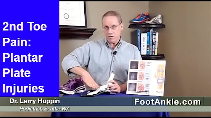

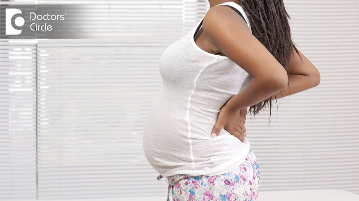

Copyright © 2024 boxhoidap Inc.